Download
1 / 4
300 likes | 2.26k Views
Neutrophil. PMN-Polymorphonuclear Leucocytes. 60-70% WBC Appearance: pink granules in cytoplasm, nucleus has 3-4 lobes Function: Phagocytosis of bacteria Azurophilic (1°) granules are "lysosomes of PMNs", occur in all leukocytes. Eosinophil (Eos). Bilobed nucleus 2-4% of WBC

E N D
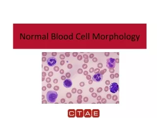
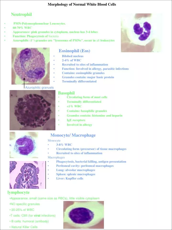
Neutrophil • PMN-Polymorphonuclear Leucocytes. • 60-70% WBC • Appearance: pink granules in cytoplasm, nucleus has 3-4 lobes • Function: Phagocytosis of bacteria • Azurophilic (1°) granules are "lysosomes of PMNs", occur in all leukocytes Eosinophil (Eos) • Bilobed nucleus • 2-4% of WBC • Recruited to sites of inflammation • Function: Involved in allergy, parasitic infections • Contains: eosinophilic granules • Granules contain: major basic protein • Terminally differentiated Morphology of Normal White Blood Cells Azurophilic granuels Basophil • Circulating form of mast cells • Terminally differentiated • <1% WBC • Contains: basophilic granules • Granules contain: histamine and heparin • IgE receptors • Involved in allergy Monocyte/ Macrophage Monocyte • 3-8% WBC • Circulating form (precursor) of tissue macrophages • Recruited to sites of inflammation Macrophages • Phagocytosis, bacterial killing, antigen presentation • Peritoneal cavity: peritoneal macrophages • Lung: alveolar macrophages • Spleen: splenic macrophages • Liver: Kupffer cells lymphocyte • Appearance: small (same size as RBCs), little visible cytoplasm • NO specific granules • 20-25% of WBC • T cells: CMI (for viral infections) • B cells: humoral (antibody) • Natural Killer Cells
Granulocyte Development • Trends: • Immature Mature • Large cell Small cell • No granules Azurophilic (non-specific) granules Cell-specific granules • Round nucleus indented nucleus U-shaped multilobed (specific for cell type) myeloblast • 15-20 m • large, euchromatic, spherical nucleus (>3 nucleoli) • basophilic cytoplasm with no granules • prominent nucleoli • can be seen in peripheral blood with certain leukemias
nucleoli Eosinophil myelocyte golgi Neutraphil myelocyte Azurophilic granuels Late Myelocyte/ Early Metamyelocyte Myelocyte (M1) promyelocyte • Spherical nucleus • Becomes increasingly heterochromatic • Prominent Golgi apparatus • Negative image • Lots of azurophilic granules • Formation of specific granules • Emerge from Golgi (cis face) complex • Characteristic staining reactions for each line • Last stage that can do mitosis • First recognizable cell in granulopoiesis -Cannot tell what kind of cell it will become • 17-26 um in diameter • Largest cell in series • Large oval nucleus • Muliple nucleoli • Golgi Ghost • Azurophilic (primary) granules in cytoplasm -Produced only at this stage
Metamyelocyte (M2) Eosinophil metamylocyte Neutraphilic metamylocytes Late neutraphilic metamylocyte • First stage that is clearly divided into separate lines • Few hundred granules present in the cytoplasm • Specific granules outnumber the azurophlic granules 4:1 • Nucleus • Heterochromatic • Indentation deepens to form horse-shoe shape Band Cells (M3) • Last immature stage in Neutrophilic series • Sometimes seen in circulation • Particularly during states of chronic infection • Nucleus is elongated and of uniform width • Nucleus constricts • 2-5 lobes are formed • PMNs