Microarray Experiment Design and Data Interpretation
Microarray Experiment Design and Data Interpretation. Susan Hester, Ph.D. Environmental Carcinogenesis Division Toxicogenomic Core Facility US EPA hester.susan@epa.gov 919-541-1320. Presentation Outline.


Microarray Experiment Design and Data Interpretation
E N D
Presentation Transcript
Microarray Experiment Design and Data Interpretation Susan Hester, Ph.D. Environmental Carcinogenesis Division Toxicogenomic Core Facility US EPA hester.susan@epa.gov 919-541-1320
Presentation Outline • Traditional biology versus genomics • Basics of genomics • Data mining goals and approaches using parallel analyses • -some examples • Interpreting changes in gene expression to identify altered molecular pathways • Evaluating pathway alterations in concert with traditional toxicology data for greater understanding of mode of action
Traditional Biology Measure one tree at a time Measure one element in 10-50 samples
“Omic” Biology Measure tens of thousands of elements in 2 to 4 samples Measure Forests (groups of trees)
Genomic research is a data-rich technology • Microarrays are called chips or arrays • Takes advantage of the natural property of DNA to pair with its complimentary strand • One strand is built into the array and then is used as a probe for the complementary strand in the biologic sample • The binding confirms the presence of mRNA or cDNA • In the sample
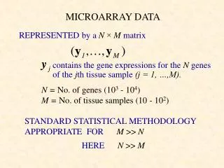
Genomic Profiling-Find ”Significantly Changed Genes” From: All probesets Typical experiment is ~ 1M datapoints To: Reduce to a much smaller number of “meaningful genes”
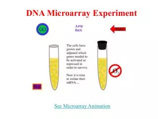
Finding genes in samples-1st step 1 genechip cell location 1 genechip apply sample
2nd step Tagged DNA fragments that base pair will glow 2nd step shine light final image text file with gene intensities
Experimental Design • Use adequate controls • Sample collection • Choose time-points and doses • Hybridization schemes-1 or 2 colors
Data Quality and Data Mining • RNA quality • Scans • Summary statistics
RNA quality: • Agilent 2100 Bioanalyzer • Measure RNA quality and quantity • Uses small sample size and take minutes Good Quality RNA Degraded RNA Agilent Gel Image
QC Assessment of Scanned Slide • Showing Good • Dynamic Range • of Signal Intensity • Low background • signal Poor scan Good scan
Summary Statistics for each array Raw gene intensity distribution for each array After normalization shows reduced variance max median min Grp 1 2 3 4 5 6
Example of with-in group outliers Example of 2 array outliers (high and low median values) Arrays
Goals of Data Mining • Reduce the large dataset by first exclude “unchanging • genes” • Early microarray papers used a simple “fold change” • to find differences • Most analyses now rely on statistical tests to identify • changed genes-supervised versus unsupervised • Find genes that distinguish the various biologic classes • “significant genes”
General Approach: From many genes to a few 28,000 rat genes 34,000 mouse genes normalize data to compare across arrays analysis begins here supervised (prior knowledge) and unsupervised (no prior knowledge) T test, ANOVA, etc. PCA, KNN, clustering genes…now associate with gene name using databases to assign gene function characterize genes into pathways explore pathways by combining into networks
Array Image Inspection Confirms the Induction of Many Genes 1 uM As50 uM As
Statistical Filter shows more significant genes at higher doses 1 uM As 50 uM As genes that have values>1.5 fold and significant p<0.05
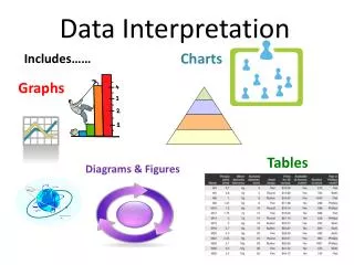
Many Views of the Data • Table of filtered genes • Principal Component Analysis (PCA) • Venn Diagrams-gene level • Correlate Transcription with Functional Assays • Map genes to pathways • Venn Diagram-pathway level
Table view: Significantly Altered Genes by Chemical, Day and Dose in rat liver
Principal Component Analysis • Identifies dose-response, if present • Assess experiment • Worth analyzing ? • Identify outliers-bad chips • Find samples with similar expression patterns What it does What it looks like: • uses all samples and genes • using statistics, reduces and plots the data • helps visualize data in 2 or 3 planes (3D) What it tells • groups samples or genes with similar profiles • differentiates treatment or exposure groups
Numbers of Common and Unique Genes Over Time (High Dose)-rat liver
Dose response corresponds to functional assays Functional assays Better description of dose response by genomics
Pathway Venn Unique and common pathways over time
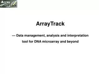
Pathway and network visualizations • cellular • molecular • network • metabolic • transcription
Example of a molecular pathway with gene intensity values added Oxidative Phosphorylation pathway red=gene induced green=gene repressed rainbow=mixed ATPase Oxidoreductase NADH dehydrogenase succinate dehydrogenase complex cytochrome c oxidase subunit
Cellular pathway extracellular cytoplasmic Note c-Jun JNK1, ERK1 repression* nuclear Expression legend Green= decreased Red=increased Rainbow=mixed
Gene Network: One Transcription factor:
Conclusions • Steps for a successful microarray experiment: • Experiment design-focus your research question • Data quality assessment • Supervised and unsupervised analyses • Integrating gene expression results with other • phenotypic endpoints