Download
1 / 36
370 likes | 574 Views
Breast Imaging Made Brief and Simple. Jane Clayton MD Associate Professor Department of Radiology LSUHSC New Orleans, LA. Breast Imaging Made Brief and Simple. What women are referred for breast imaging?

E N D
Breast Imaging Made Brief and Simple Jane Clayton MD Associate Professor Department of Radiology LSUHSC New Orleans, LA
Breast Imaging Made Brief and Simple What women are referred for breast imaging? Two groups of women are referred for breast imaging, those without symptoms (asymptomatic) and those with symptoms.
Breast Imaging Made Brief and Simple The asymptomatic women have a screening mammogram to look for small nonpalpable abnormalities in the breasts.
Breast Imaging Made Brief and Simple CC 60 yo asymptomatic
Breast Imaging Made Brief and Simple MLO 60 yo asymptomatic
Breast Imaging Made Brief and Simple Irregular shaped mass with indistinct margins on the left at 8:00 in the middepth of the breast BI-RADS 4 Biopsy recommended Dx: Invasive ductal carcinoma
Breast Imaging Made Brief and Simple BI-RADS is an assessment scale indicating the likelihood of breast cancer for mammographic findings.
Breast Imaging Made Brief and Simple • 0 Further information needed to put in assessment category • 1 Normal • 2 Benign finding • 3 Probably benign-6 mo followup • 4 Suspicious-biopsy • 5 Malignant-biopsy
Breast Imaging Made Brief and Simple The women with symptoms have a diagnostic mammogram first. Symptoms usually include a palpable mass or nipple discharge.
Breast Imaging Made Brief and Simple The area of concern is marked with a BB. BB indicating palpable mass
Breast Imaging Made Brief and Simple Further imaging workup for these women includes additional mammographic views and ultrasound.
Breast Imaging Made Brief and Simple Breast Cancer Statistics • Most common malignancy in American women (except skin) • Approximately one third of new cancers diagnosed • Second leading cause of death from cancer • 211,300 new cases of invasive cancer this year • 55,700 new cases of DCIS this year • Leading cause of premature mortality- average 18.5 potential years of life lost
Breast Imaging Made Brief and Simple Breast Cancer Statistics If breast cancer is diagnosed while the disease is local survival is 96%. Survival for regional disease is 78%. Survival for distant disease is 21%.
Breast Imaging Made Brief and Simple Methods of detection of breast cancer: Breast Self Examination Clinical Breast Exam Mammography
Breast Imaging Made Brief and Simple Methods of Detection of Breast Cancer Breast Self Examination and Clinical Breast Examination are used in the women under 40 yo to detect palpable masses.
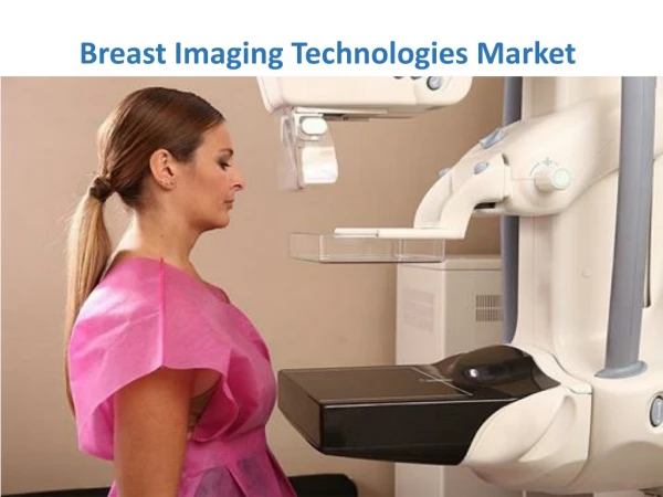
Breast Imaging Made Brief and Simple Methods of Detection of Breast Cancer At 40 and older mammography is used to screen for breast cancer in women without symptoms. Mammography is the most sensitive examination for detection of early breast cancers.
Breast Imaging Made Brief and Simple Methods of detection of breast cancer At 40 and older Breast Self Examination and Clinical Breast Examination are used to detect breast cancers not seen on a mammogram because of technical limitations, interval tumor growth or breast cancers missed on the mammogram.
Breast Imaging Made Brief and Simple American Cancer Recommendations for Screening Mammography Begin screening at age 40 unless the woman has a mother or sister who developed breast cancer before menopause. Screen annually. No end age for screening mammography.
Breast Imaging Made Brief and Simple Risk factors for development of breast cancer Female Age > 35 Early menarche Late menopause Nulliparity Pregnancy after 30 Affected first degree relative (mother, sister, daughter) Previous history of breast cancer Biopsy proof of atypical epithelial proliferation Biopsy proof of lobular carcinoma in situ
Breast Imaging Made Brief and Simple The reduction in mortality for women whose breast cancers were detected on a screening mammogram is 30% or higher.
Breast Imaging Made Brief and Simple Mammographic findings of breast cancer Mass Microcalcifications Mass and microcalcifications
Breast Imaging Made Brief and Simple Mammographic findings of breast cancer Mass
Breast Imaging Made Brief and Simple Asymptomatic screening mammogram
Breast Imaging Made Brief and Simple Diagnosis: 1.2 cm invasive ductal carcinoma with associated low grade DCIS
Breast Imaging Made Brief and Simple Mammographic findings of breast cancer Microcalcifications
Breast Imaging Made Brief and Simple Mammographic findings of breast cancer Mass and microcalcifications