Clinical Case
250 likes | 1.24k Views
3º CURSO DE DOENÇAS PULMONARES DIFUSAS. Clinical Case. February 2014. Vanessa Santos , Susana Guimarães , Conceição Souto Moura, José Miguel Jesus, Patrícia Mota, Natália Melo, António Morais. IDENTIFICATION. MCMC Female 53 years old Caucasian

Clinical Case
E N D
Presentation Transcript
3º CURSO DE DOENÇAS PULMONARES DIFUSAS Clinical Case February 2014 Vanessa Santos, Susana Guimarães, Conceição Souto Moura, José Miguel Jesus, Patrícia Mota, Natália Melo, António Morais
IDENTIFICATION • MCMC • Female • 53 years old • Caucasian • Works as a housewife, worked in textile factory for 24 years 3º Curso de Doenças Pulmonares Difusas
PREVIOUS MEDICAL HISTORY • Recurrent otitis in childhood • No other relevant pathological antecedents • No surgical history • No usual medication • No allergies • No smoking history • No drugs or alcohol consumption 3º Curso de Doenças Pulmonares Difusas
PREVIOUS MEDICAL HISTORY • No contact with birds or other animals • No recent travelling abroad • No history of contact with tuberculosis • No sexual risk behaviours • No history of blood transfusions 3º Curso de Doenças Pulmonares Difusas
HISTORY OF PRESENT ILLNESS Previously asymptomatic • 2 MONTHS HISTORY OF: • Dyspnoea on exertion • Asthenia, anorexia • Weight loss (4Kg) • SINCE TWO WEEKS AGO: • Fever 20 1 3 She visited her primary care physician treated with antibiotics without clinical improvement.
HISTORY OF PRESENT ILLNESS Persistence and worsening of the symptoms HOSPITAL • Pale. Hydrated. • Febrile (temperature 38.4ºC). • Normal blood pressure. Tachycardia (125 beats per minute). • No digital clubbing. • No respiratory distress. SpO2 (FiO2 21%): 98% • Cardiac and pulmonary auscultation with no abnormalities. ON A DM I S S I ON 3º Curso de Doenças Pulmonares Difusas
DIAGNOSTIC TEST RESULTS • BLOOD ANALYSIS • Hypocapnia • Anaemia • Hyperleukocytosis • Thrombocytopenia • CRP elevated 3º Curso de Doenças Pulmonares Difusas
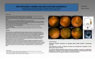
DIAGNOSTIC TEST RESULTS • CHEST X-Ray • Strengthening of bilateral pulmonary hila • Without opacifications 3º Curso de Doenças Pulmonares Difusas
DIAGNOSTIC TEST RESULTS • MYELOGRAM Hypercellular bone marrow considering the age of patient. Scarce megakaryocytes. Infiltration by a monomorphic population of immature cellsof medium size, high ratio n/c, lax chromatin, nucleus with 2-3 visible nucleoli, scant agranular cytoplasm and one Auer’s body. Scarce myeloid differentiation. Scant representation of the erythroid series. Differential (300 cells): Bla 92%, PMY 0%, My 0%, M&Bands 0.66%, Neut 1%, Eos 0%, Mon 0.33%, Lym 5.33%, Plas 0%, Erythro 0.66%. M/E Ratio 10:1. IFT: 95% blast cells of the myeloid line: CD33+, CD117+ (62%), CD56+ (40%), CD64+ (39%), CD123+, CD34-, CD13-, CD11b-, CD15-CD16-, CD2-, CD36-, CD6-, HLADr- 3º Curso de Doenças Pulmonares Difusas
DIAGNOSTIC TEST RESULTS • MYELOGRAM • Hypercellular bone marrow for age. Scarce megakaryocytes. • Massive infiltration monomorphic population of immature cells of medium size, high aspect ratio n/c, lax chromatin, nucleus with 2-3 visible nucleoli, scant cytoplasm and agranular, with various extensions and 1body Auer. Scarce myeloid differentiation. Sparse representation of the erythroid series. • Differential (300 cells): Bla 92%, PMY 0%, My 0%, M&Bands 0.66%, Neut 1%, Eos 0%, Mon 0.33%, Lym 5.33%, Plas 0%, Erythro 0.66%. M/E Ratio 10:1. • IFT: 95% blast cells of the myeloid line: CD33+, CD117+(62%), CD56+(40%), CD64+(39%), CD123+, CD34-, CD13-, CD11b-, CD15-CD16-, CD2-, CD36-, CD6 -, HLADr- • ACUTE MYELOID LEUKAEMIA, M1 FAB • Cytapheresis and Hydroxycarbamide • Started induction protocol AML-12 on 22/06/2013 with: • Cytarabine • Daunorubicin • Etoposide With clinical and analytic improvement 3º Curso de Doenças Pulmonares Difusas
EVOLUTION 28 DAYS AFTER HOSPITALIZATION: • Worsening of general condition • Dyspnoea on minimal exertion • Dry cough • Chest pain • Fever again • SpO2 (FiO2 21%): 96% • Pulmonary auscultation: inspiratory crackles on the right • No increase in inflammatory parameters • No microbiological isolation • Echocardiogram showed no valvular vegetations 3º Curso de Doenças Pulmonares Difusas
DIAGNOSTIC TEST RESULTS • CHEST X-RAY – Bilateral patchy and heterogeneous, not well defined opacifications, mainly in the right lung. Previous 3º Curso de Doenças Pulmonares Difusas
DIAGNOSTIC TEST RESULTS • HIGH-RESOLUTION CHEST COMPUTED TOMOGRAPHY • Bilateral pulmonary opacifications with ground glass halo, predominantly peripheral distribution • Some patchy ground glass opacities • Interlobular septal thickening • Bilateral pleural effusion 3º Curso de Doenças Pulmonares Difusas
DIAGNOSTIC TEST RESULTS • FLEXIBLE BRONCHOSCOPY • No endobronchial lesions • Bronchial lavage (BL) fluid specimens were smear and culture negative for common bacteria and acid-bacilli. BL was also negative for the markers of other infections. 3º Curso de Doenças Pulmonares Difusas
EVOLUTION ANTIBIOTICS were initiated because she kept symptoms and had radiological alterations, even considering that no increase in inflammatory parameters and no microbiological isolation were detected • Without clinical improvements CT – GUIDED TRANSTHORACIC LUNG BIOPSY 3º Curso de Doenças Pulmonares Difusas
Prominent proteinaceous exudation within alveolar spaces; some inflammatory cells.
DIAGNOSTIC TEST RESULTS • CT – GUIDED TRANSTHORACIC LUNG BIOPSY • Fibrous and proteinaceous exudates with some signs of organization, associated with inflammatory cells in the alveolar spaces. • Interstitial lymphocytic inflammatory infiltrate, fibrosis and hyperplasia of pneumocytes. • No hyaline membranes. No eosinophils. • No signs of infection. No signs of malignancy. • PAS and Grocott methods were negative. • Microbiological exam: negative. 3º Curso de Doenças Pulmonares Difusas
DIAGNOSTIC TEST RESULTS • CT – GUIDED TRANSTHORACIC LUNG BIOPSY • Fibrinoid and proteinaceous exudates associated with inflammatory cell in the alveolar spaces. • Interstitial lymphocytic inflammatory infiltrate, fibrosis and hyperplasia of pneumocytes. • No signs of infection. • No signs of malignancy. • PAS and Grocott methods were negative. ACUTE FIBRINOUS AND ORGANIZING PNEUMONIA 3º Curso de Doenças Pulmonares Difusas
SUMMARY • Dyspnoea on exertion, asthenia, anorexia and weight loss since two months ago and then fever. The patient was admitted in the hospital due to complaints and analytic alterations. • During hospitalization was diagnosed Acute Myeloid Leukaemiaand treated withcytapheresis, hydroxycarbamide, cytarabine, daunorubicin and etoposide. • 28 days after hospitalization and 24 days after starting chemotherapy, a worsening of general condition was detected, with dyspnoea on minimal exertion, dry cough and fever again. No microbiological isolation. • Thoracic CT: Bilateral pulmonary opacifications with ground glass halo, predominantly peripheral distribution. Some patchy ground glass opacities. Interlobular septal thickening. Pleural effusion. • CT-guided lung biopsy: Acute Fibrinous and Organizing Pneumonia (AFOP)
TREATMENT ACUTE FIBRINOUS AND ORGANIZING PNEUMONIA • Prednisolone 40 mg/day Weight: 50 Kg Later... Reduction of corticosteroid therapy (prednisolone 20 mg/day) and treatment with azithromycin 3x/week was suggested due to haematological disease, the infection risk and the possibility of bone marrow transplantation. 3º Curso de Doenças Pulmonares Difusas
EVOLUTION 1 month after diagnosis Improvement of respiratory complaints • HRCT: • Pneumatoceles • Some areas with tenuous ground glass opacities Previous 3º Curso de Doenças Pulmonares Difusas
EVOLUTION 2 months after diagnosis No respiratory complaints • HRCT: • Improvement of radiological changes Previous 3º Curso de Doenças Pulmonares Difusas
EVOLUTION No dyspnoea on exertion. No cough or sputum. No signs of respiratory distress. 6 months after diagnosis Chest X-Ray and HRCT: without opacifications ABG: pH 7.41, pO2 99.7, pCO2 38.2, HCO3- 23.7, SpO2 96.4% 3º Curso de Doenças Pulmonares Difusas
? TO BE CONSIDERED • AFOP due to altered immune status of acute leukaemia? • AFOP due to acute leukaemia treatment? • Other aetiologies? • Which treatment and how long? • Considering the haematological disease, which dose of corticoid can we use? 3º Curso de Doenças Pulmonares Difusas