Download
1 / 135
1.36k likes | 1.5k Views
Cells. I. Basic History. Every. _______ living thing, from the tiniest bacterium to the largest whale, are made of one or more cells!

E N D
I. Basic History Every • _______ living thing, from the tiniest bacterium to the largest whale, are made of one or more cells! • Before the seventeenth century, no one knew that _______ existed. CELLS
I. Basic History small unaided eye • Most cells are too _______ to be seen with the _____________. • Not discovered until after the invention of the ____________ in the early 17th century. microscope
II. Important Scientists Anton von Leeuwenhoek FIRST OBSERVE • A Dutch drapery storeowner ________________________, became the _______ person to _________ and __________ MICROSCOPIC ORGANISMS and LIVING CELLS. DESCRIBE
II. Important Scientists Robert Hooke • 1665: the English scientist _______________ used a microscope to examine a thin slice of ______ and described it as consisting of “a great many little boxes”. It was after his observation that Hooke called what he saw “_______”. They looked like “little boxes” and reminded him of the small rooms in which monks lived. So he called them “_______”. cork cells cells
II. Important Scientists plant animal • 1824: the French scientist Henri Dutrochet, concluded that ______ and ________ tissue were always made up of cells. • 1831: Robert Brown named the _________ nucleus
II. Important Scientists • 1838: German botanist Matthias Schleiden concluded that all ________ are made of cells. • 1839: German zoologist Theodor Schwann reported that _________ are also made of cells. plants animals
II. Important Scientists • 1845: Felix Dujardin studied the living cell and noted it contained a material called _____________. • 1855: German physician Rudolf Virchow induced that ALL cells come from _____________ cells. protoplasm preexisting
II. Important Scientists • The COMBINED work of Schleiden, Schwann, and Virchow makeup what is now known as the modern _____________. cell theory
III. The Cell Theory Consists of 3 Principles composed cells • All living things are ____________ of one or more _______. • _______ are the basic units of ___________ and __________ in an organism. • Cells come ______ from the _______________ of __________ cells. Cells structure function ONLY existing reproduction
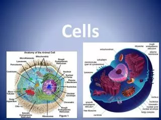
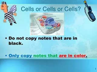
IV. Two Types of Cells EUKARYOTE other nucleus • _______________ = cell that contains a _________ and _______ ___________ ____________________ Ex: ________, fish, mammals, _________ and ________ membrane-bound organelles plants insects humans
IV. Two Types of Cells PROKARYOTE lacks nucleus other • _______________ = cell that _______ a _________ and _____ ___________________ __________. Ex: _____________ organisms such as __________ and their relatives membrane-bound organelles unicellular bacteria
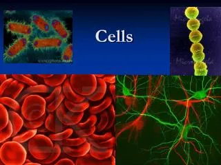
V. Cell Diversity alike • Not all cells are _______. • Cells within the same organism show enormous diversity in ____, ______, and ____________________. • Your body contains at least _____ different cell types! size shape internal organization 200
VI. Cell Size: unaided • A few types of cells are large enough to be seen by the _________ eye. • _______________ is the _________ cell in the body and can be seen without the aid of a microscope. • Most cells are visible only with a ____________. Female egg largest microscope
VI. Cell Size: Most cells are small for 2 reasons RATIO • ________________________________: • Cells are limited in size by the _______ between their _____________________ and their ________. • As a cell’s size increases, its volume increases much faster than its surface area. • (see picture on the next slide!) outer surface area volume
VI. Cell Size: • The cell’s nucleus (the brain) can only control a certain amount of living, active cytoplasm.
VII. Cell Shape: Variety • _________ of shapes • The _______ of the cell depends on the __________. shape function
VII. Cell Shape: Nerve cells • Ex: ____________ that carry information from your toes to your brain are long and threadlike. • Ex: ____________ are shaped like round discs that can squeeze through tiny blood vessels. Blood cells
VIII: Cellular Organization Multicellular organisms • _______________________ are made up of many cells, each of which is specialized to perform a distinct function. • Digestion, movement, respiration, filtering, etc. • __________________ DO NOT carry out ALL life functions, but rather depend on each other. Individual cells
VIII: Cellular Organization Tissue • ________ = a group of cells functioning together to perform an activity. • Ex: muscle and nerve tissues • Ex: Plant tissues = stem and root • ________ = groups of two or more tissues that function together. • Stomach, leaf of a plant • Cooperation among organs makes life functions within an organism efficient. Organs
VIII: Cellular Organization Tissue • ________ = a group of cells functioning together to perform an activity. • Ex: muscle and nerve tissues • Ex: Plant tissues = stem and root • ________ = groups of two or more tissues that function together. • Stomach, leaf of a plant • Cooperation among organs makes life functions within an organism efficient. Organs
VIII: Cellular Organization Summary Cells Tissues Organs
Always carry a microscope with one hand holding the arm and one had under the base.
What’s my Power? power multiply ocular lens power • To calculate the power of magnification, __________ the _______ of the _____________ by the _______ of the ____________. objective O Ocular lens (10X) Objectives (4X, 10X, 40X) O O
What’s my Power? • Low Power • Ocular lens = 10X • Objective = 4X • TOTAL magnification for LOW power = _________ 40X
What’s my Power? • Medium Power • Ocular lens = 10X • Objective = 10X • TOTAL magnification for MEDIUM power = _________ 100X
What’s my Power? • High Power • Ocular lens = 10X • Objective = 40X • TOTAL magnification for HIGH power = _________ 400X
Comparing Powers of Magnification: HIGHER • We can see better details with ________ powers of magnification, but we can’t see as much of the image.
higher power • Which of these images would be viewed at a ______________ of magnification?
Compound Light Microscopes • You will be using a compound light microscope in several labs. • These microscopes have a maximum magnification of 400X • So you __________ see most of the organelles like ribosomes, Golgi bodies, lysosomes, etc. • More powerful microscopes are needed (2000X plus) CANNOT
Common Problem . . . AIR BUBBLES AIR BUBBLES AIR BUBBLES
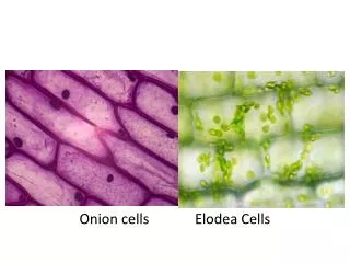
Stained Onion Cells • Can you identify the cell walls? • Can you identify any other organelles?
Elodea – Typical Plant Cells as seen with the light microscope
How to Make a Wet-Mount Slide 1. Get a clean slide and cover slip from your teacher. 2. Place ONE drop of water/iodine in the middle of the slide. Don’t use too much or the water will run off the edge and make a mess! 3. Place the edge of the cover slip on one side of the water/iodine drop. 4. Slowly lower the cover slip on top of the drop. 5. Place the slide on the stage and view it first with the LOW power objective. Once you see the image, you can rotate the nosepiece to view the slide with the different objectives.
Let’s give it a try . . . • Turn on the microscope and then rotate the nosepiece to click the LOW power objective into place. • Place a slide on the stage and secure it using the stage clips. Use the coarse adjustment knob (large knob) to get the image into view and then use the fine adjustment knob (small knob) to make it clearer. • Once you have the image in view, rotate the nosepiece to view it under different powers. Draw what you see on a piece of paper. BE CAREFUL WITH THE LARGEST OBJECTIVE! Sometimes there is not enough room and you will not be able to use it!