Download
1 / 27
300 likes | 679 Views
Gastrointestinal pathogens. Vibrio V. cholerae : cholera V. parahaemolyticus : gastroenteritis V. vulnificus : wound infection; primary sepsis Aeromonas A. hydrophila : gastroenteritis; wound infection Campylobacter C. jejuni : gastroenteritis C. fetus : septicemia Helicobacter

E N D
Gastrointestinal pathogens Vibrio V. cholerae: cholera V. parahaemolyticus: gastroenteritis V. vulnificus: wound infection; primary sepsis Aeromonas A. hydrophila: gastroenteritis; wound infection Campylobacter C. jejuni: gastroenteritis C. fetus: septicemia Helicobacter H. pylori: gastritis; peptic ulcer; gastric cancer, MALToma
Vibrio Gram-negative curved rod. Move rapidly by means of a polar flagellum. Grow at 14oC to 40 oC on many kinds of media. Grow at a high pH and are rapidly killed by gastric acid. Oxidase-positive. Vibrio species inhabit estuarine and marine environments, and are either halotolerant or halophilic (this property differentiates Vibrio from Aeromonas).
V. cholerae Antigenic structure and biological classification O antigen: confers serological specificity (more than 140 O serogroups). Serogroups O1 andO139 cause cholera; some strains of the other serogroups may cause cholera-like disease. Two biotypes of V. cholerae O1: classical and El Tor. HemolysinV-P testPolymixin B classical -- sensitive El Tor + + resistant
V. cholerae Pathogenesis and Immunity Under natural conditions, V. cholerae is pathogenic only for humans. Mean infective dose: 108-1010. Gastric acid provides some protection. Mouth intestine attach to the microvilli of the epithelial cells and multiply release cholera toxin. Major virulence factors: Toxin-coregulated pili (TCP): adherence to mucosal cells. Enterotoxin (cholera toxin): produced by O1 and O139 strains. Hemagglutinin-protease: releases bacteria from mucosal cells. other enterotoxins, flagellum, siderophores.
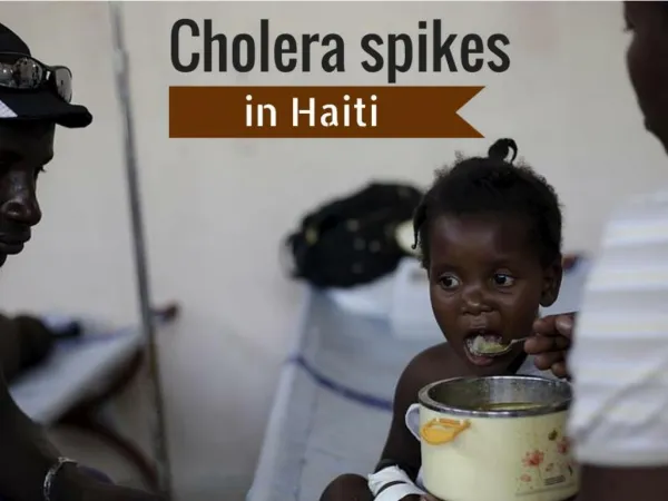
V. cholerae Clinical Diseases In many instances only 1-5% of exposed susceptible persons develop disease. Incubation period: 1-4 days. Sudden onset of nausea and vomiting, and profuse diarrhea with abdominal cramps; "rice water" stool (containing mucus, epithelial cells, and large numbers of vibrios) rapid loss of fluid and electrolytes profound dehydration that leads to circulatory collapse (hypovolemic shock). Carrier state seldom exceeds 3-4 weeks, and true chronic carriers are rare. The El Tor biotype tends to cause milder disease than the classical biotype.
V. cholerae Treatment Water and electrolyte replacement - most important. Antibiotic treatment: tetracycline or trimethoprim-sulfamethoxazole. Diagnostic lab tests Specimens: mucus flecks from stool. Smears: Dark-field or phase contrast microscopy may show the rapidly motile vibrios. Culture: peptone agar, blood agar (pH near 9.0), or TCBS (thiosulfate-citrate-bile salts-sucrose) agar. Alkaline peptone broth can be used for enrichment. Biochemical tests Serological tests: slide agglutination tests using anti-O1 or O139 antiserum.
V. cholerae • Epidemiology, Prevention, and Control • Seven pandemics since 1817 • classical biotype: till early 1960s • El Tor: emerging in Asia in 1961 • New epidemic: 1992, India, V. cholerae O139 Bengal. • Endemic • Natural reservoir: estuarine and marine environments. • Transmission: • water, food, and flies • Controls: • Improvement of sanitation and personal hygiene. • Isolation of patients, and their excreta be disinfected. • Vaccination: in development.
內急>_<!台南市出現第二例本土性霍亂! 「台南巿疑似出現國內第二例本土性霍亂病例,請民眾吃熟食並注意個人衛生」 疾管局防疫組於 2005/7/5 發布 疾病管制局於今年6月28日下午接獲台南縣某醫院以「腹瀉症候群」進行通報(女性,78歲,居住於台南巿,發病前曾因肺癌接受化療,抵抗力較差),該局於7/3初步由個案糞便檢體已檢出霍亂弧菌(O1小川型),該菌株正進行產毒性試驗。疾管局立即與台南巿衛生局共同進行疫調、防疫工作(含衛教、住家環境消毒、接觸者及環境檢體採檢等),並依規定將個案予以隔離治療。該個案密切接觸者(6人)及住家環境檢體(浴室洗手檯、廚房洗手檯、砧板、菜刀,共4件),檢出結果皆為陰性。由於該個案發病日(6/24)與日前臺南縣柳營鄉今年本土性首例霍亂個案發病日(6/16)相近,初步疫調未發現兩者有關聯性,但疾管局為求慎重,仍將該兩者個案檢體進行菌株基因比對,期能釐清案情。目前該個案病況已改善,疫調結果尚未發現其他疑似個案,衛生單位仍將持續監控。
V. parahaemolyticus Halophilic Causes acute gastroenteritis following ingestion of contaminated seafood (e.g., raw fish or shellfish), and wound infections rarely. Incubation period: 5-72 hours. Symptoms: sudden onset of nausea, vomiting, abdominal cramps, no or low grade fever, and watery (mostly) to bloody diarrhea. Disease subsides spontaneously in 1-4 days. Most virulent strains produce a thermostable hemolysin (TDH; also called Kanagawa hemolysin), which is enterotoxic (by inducing chloride ion secretion), cytotoxic and cardiotoxic in experimental animals.
V. vulnificus Halophilic. Capsular polysaccharides are the major virulence factor. Causes sporadic cases of two forms of disease: Wound infections (initial swelling, erythema, and pain followed by vesicles or bullae and eventually tissue necrosis) with or without secondary septicemia. Primary septicemia with or without skin lesions after ingestion of undercooked seafood (particularly raw oysters). Mortality rate can be as high as 50% without prompt antibiotic treatment and surgical debridement. Most patients of V. vulnificus infection have underlying conditions such as chronic liver diseases, alcoholism, diabetes mellitus, steroid administration as well as other chronic diseases.
Aeromonas Gram-negative, facultative anaerobic bacilli. Morphologically resembles Enterobacteriaceae; oxidase-positive. The most important pathogen: A. hydrophila. Ubiquitous in fresh and brackish water. Diseases associated with Aeromonas: Opportunistic systemic disease, particularly in those with hepatobiliary disease or an underlying malignancy. Acute watery or dysenteric diarrhea (may become chronic) by congestion of contaminated water or food. Wound infections acquired by exposure to contaminated water. Numerous potential virulence factors have been proposed, however, their precise roles are unknown. Resistant to multiple drugs.
Campylobacter C. jejuni, C. upsiliensis, C. coli: enteritis, and occasionally systemic invasion. C. fetus: opportunistic infections (septicemia, meningitis, etc.) Campylobacters contain pathogens for various animals, which serve as reservoirs, and may cause sepsis, abortion, or enteritis (Zoonotic). C. jejuni is a common cause of diarrhea in humans. Morphology Small gram-negative rods with coma, S or “gull wing" shapes. Motile with a single polar flagellum.
Campylobacter Physiology and Structure Can be classified by the O antigens, capsular antigens and flagellar antigens. Microaerophilic: grows best in the presence of 5% O2 + 10% CO2 (anaerobic jar without catalyst plus gas generating pack or gas exchange). Most Campylobacter pathogens (except for C. fetus) can grow at 42 oC (C. jejuni grows better at 42 oC than at 37 oC). Oxidase- and catalase-positive. Do not ferment or oxidize carbohydrates.
Campylobacter Pathogenesis and Clinical diseases C. jejuni Infections are acquired from ingestion of contaminated food, particularly, poultry. Roles of potential virulence factors, e.g., adhesins, cytotoxins, and enterotoxins, remain unknown because of lack of an animal model. Mouth multiply in the small intestine invade the epithelium cause inflammation presence of RBC and WBC in the stools. Ingestion of about 104 bacteria is usually necessary to produce infection.
Campylobacter Pathogenesis and Clinical Diseases C. jejuni Acute onset of crampy abdominal pain, profuse diarrhea that may be grossly bloody, headache, malaise, and fever. Usually self-limited (5-8 days). Occasionally, the bloodstream is invaded and a clinical picture of enteric fever develop. Guillain-Barré syndromeis an autoimmune disease of the peripheral nervous system resulting from infection by C. jejuni and C. upsaliensis due to antigenic cross-reactivity between lipopolysaccharide and glycosphingolipids of the neural tissue in the peripheral nervous system. Reactive arthritis is another immune-related complication of campylobacter infections.
Campylobacter Pathogenesis and Clinical Diseases C. fetus Opportunistic pathogen causing systemic infections in debilitated and immunocompromised patients. In most cases, the patients develop gastroenteritis first, and then septicemia with dissemination to multiple organs. This organism is more resistant to complement- and antibody-mediated serum killing than other Campylobacter pathogens. This is mediated by a surface protein, S protein, that forms a capsule-like structure which may inhibit C3b binding to the bacteria.
Campylobacter Laboratory Diagnosis Specimens: diarrheal stool Smears: small, gram-negative "gull-wing" shaped rods. Commercial kits for detection of specific antigens in the specimen. Culture: selective media (e.g., Skirrow's medium and Camp BAP medium, in which blood, charcoal, and antibiotics are added), microaerophilic environment; 42 oC for C. jejuni. Treatment, Prevention, and Control Replacement of lost fluid and electrolytes. Antibiotics used for severe infections or septicemia. Prevention: proper preparation of food; avoidance of unpasteurized dairy products; preventing contamination of water supplies.
Helicobacter H. pylori: gastritis, peptic ulcer, gastric adenocarcinoma, gastric mucosa-associated lymphoid type (MALT) B-cell lymphomas. Other helicobacters may cause gastroenteritis and proctitis. Morphology and Physiology Spiral-shaped, gram-negative rod. Highly motile (corkscrew motility) with polar flagella. Grow on complex media in microaerophilic (5% O2 and 10% CO2 ) conditions. Do not ferment or oxidize carbohydrates.
H. pylori Physiology Grows optimally at a pH of 6.0-7.0. Able to survive in the stomach because 1. it stays deep in the mucus layer near the epithelial surface where physiological pH is present; 2. it produces a potent urease, which catalyzes production of ammonia that further neutralizes the acid; 3. It produces an acid-inhibitory protein that blocks acid secretion from parietal cells. *Urease is found in Helicobacter spp. that colonize the stomach, but not those colonizing the intestine. Dr. Barry Marshall
H. pylori Pathogenesis and Clinical Diseases Present on the gastric mucosa of less than 20% of persons under 30 but increases in prevalence to 40%-60% (over 80% in developing countries) of persons age 60. H. pylori is associated with type B gastritis, gastric and duodenal ulcer, gastric cancer, and MALT B-cell lymphomas (MALToma). Chronic gastritis is a risk factor for gastric carcinoma. Colonization with H. pylori may protect the body from gastroesophageal reflux disease and adenocarcinomas of the lower esophagus and gastric cardia.
H. pylori Pathogenesis and Clinical Diseases Hypothetical steps in ulcer formation: Colonization of the epithelial cells (inhibit gastric acid secretion, neutralization of gastric acid, pass through the gastric mucus, adhere to the epithelial cells) Localized tissue damage by urease byproducts, enzymes (mucinase, phospholipase etc.), and cytotoxins (vacuolating cytotoxin A and cytotoxin-associated antigen) induction of chemokine production and stimulation of inflammatory response by LPS, urease, etc. Release of proteases and ROS by neutrophils results in damage of mucous membrane.
A & B C D
H. pylori Laboratory diagnosis Histological examination of gastric biopsies Warthin-Starry silver stain. Culturing biopsy specimens Culture of multiple biopsy specimens is required *Urease test is a rapid methods for detection of H. pylori in biopsy specimens or pure culture. Detection of antiurease antibodies in the blood. Urea-breath test Treatment, Prevention, and Control Treatment: Bithmus plus antibiotics Omeprazole (a proton pump inhibitor), clarithromycin plus ammoxicillin or metronidazole Humans are the main reservoir. Mechanism of transmission is most likely oral-fecal, and improved hygienic standards can be helpful for prevention.
Cholera toxin An A-B toxin produced from prophage CTXF. Subunit B promotes entry of subunit A into the cell. Subunit A activates adenylate cyclase and increases intracellular levels of cAMP, resulting in prolonged hypersecretion of water and electrolytes (can be 1 L/h) watery diarrhea, dehydration, shock, acidosis, and death. Antigenically related to LT of E. coli. Stimulates production of neutralizing antibodies.
Skin lesions: initial swelling, erythema, and pain followed by the development of vesicles or bullae and, eventually, tissue necrosis