Download
1 / 17
170 likes | 273 Views
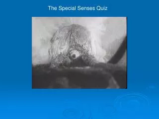
Test your knowledge on eye and ear anatomy with this quiz! Identify structures in X-rays and answer questions on canine and bovine anatomy. Learn about the zygomatic arch, lacrimal sac fossa, tympanic bulla, and more. Improve your understanding of special senses with each question in this interactive quiz.

E N D
Special Senses Imaging Quiz Developed by: SorchaMcCaughley & Mark Brims Approved by: Gawain Hammond & Maureen Bain Supported by: The Chancellor’s Fund
Special Senses Imaging Quiz START! Developed by: Sorcha McCaughley & Mark Brims Supported by: The Chancellor’s Fund
Special Senses • The Eye Q1 • The Ear Q2
The Eye Q1 Canine • (i) A makes up part of the Orbit. What is it? • Zygomatic Arch • Zygomatic Process • (ii) Which of the fossae, foraminae and fissures in the Orbit is connected, via a duct, to the nasal cavity? • Ethmoidal Foraminae • Lacrimal Sac Fossa • Orbital Fissure A Canine – Contrast showing duct in (ii).
Correct Canine • Yes! A is the Zygomatic Arch! • The Zygomatic Process is the incomplete Zygomatic Arch in horses and ruminants. • Try (ii)! • Choose a new question. Zygomatic Arch Bovine Zygomatic Process
Incorrect Canine • No, A is not the Zygomatic Process! • Remember: the Zygomatic Process is the incomplete Zygomatic Arch found in horses and ruminants. • Try again! • Choose a new question. Zygomatic Arch Bovine Zygomatic Process
Correct • Yes! The Lacrimal Sac Fossa connects with the Nasolacrimal Duct! • Try The Ear Q2! • Choose a new question. Nasolacrimal Duct
Incorrect • No, the Ethmoidal Foraminae do not connect with the nasal cavity. • Blood vessels and branches of the Ophthalmic nerve are found in the Ethmoidal Foraminae. • Try again! • Choose a new question.
Incorrect • No, the Orbital Fissure does not connect with the nasal cavity. • The Orbital Fissure carries nerves and the ophthalmic vein. • Try again! • Choose a new question.
The Ear Q2 • In this x-ray you can see the outline of the ear. • (i) Why is it not as white as the bones of the skull? • Answer. • (ii) What is A? • Tympanic Bulla • External Ear Cartilages • External Acoustic Meatus • (iii) What is B? • Tympanic Bulla • External Ear Cartilages • External Acoustic Meatus B A
Answer • Normally, cartilage cannot be seen on an x-ray as it is softer and less dense than bone. • This x-ray would be described as ‘under-exposed’. This means that the soft tissue structures are more visible. • Try (ii)! • Choose a new question.
Correct • Yes! A is the External Ear Cartilage! • It is not usually visible on x-rays as it is cartilage, not bone. • Try (iii)! • Choose a new question.
Incorrect • No, A is not the Tympanic Bulla. • The Tympanic Bullae are shown in this x-ray. • Try again! • Choose a new question. Tympanic Bulla Tympanic Bullae
Incorrect • No, A is not the External Acoustic Meatus. • The External Acoustic Meatus connects the External Ear to the Tympanic Bullae. • Try again! • Choose a new question.
Correct • Yes! B is the External Acoustic Meatus! • The outer part, nearest the external ear, is cartilagenous; it is rarely visible on x-rays. • The inner part, as it approaches the Tympanic Bulla, is osseus; it can sometimes be seen in x-rays. • Choose a new question. External Acoustic Meatus
Incorrect • No, B is not the External Ear Cartilage. • The External Ear Cartilages support the pinna of the Ear. • Try again! • Choose a new question. External Ear Cartilage
Incorrect • No, B is not the Tympanic Bulla. • The Tympanic Bulla is found at the end of the External Acoustic Meatus. • Try again! • Choose a new question. Tympanic Bulla Tympanic Bullae