Download
1 / 20
220 likes | 643 Views
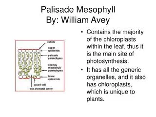
Palisade Mesophyll By: William Avey. Contains the majority of the chloroplasts within the leaf, thus it is the main site of photosynthesis. It has all the generic organelles, and it also has chloroplasts, which is unique to plants.

E N D
Palisade MesophyllBy: William Avey • Contains the majority of the chloroplasts within the leaf, thus it is the main site of photosynthesis. • It has all the generic organelles, and it also has chloroplasts, which is unique to plants.
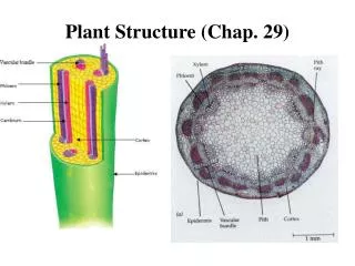
xylem: the vascular tissue in plants that conducts water and dissolved nutrients upward from the root and also helps to form the woody element in the stem. there sole purpose is to assist with the transport of water through vessels and elongated dead cells whom contain no organelles prominent organelles : xylem contains many cells with in its tissue such as tracheids, vessel elements, and parenchyma
Bundle Sheath Cell The are photosynthetic cells around the veins of a leaf. They are packed very tightly & form protection. Mitochondria & Chloroplasts are important for the structure of these cells. Chloroplasts are where the Calvin Cycle takes place. The Calvin Cycle is part of photosynthesis (production of sugar from CO2) Mitochondria are structural. Membrane folds up to cristae.
Root Hair • Absorbs water and minerals from soil and sends it to the rest of the plant • Have a large vacuole to allow more water to be absorbed
Neutrophil Neurtophil’s are the most abundant type of white blood cells in mammals. They form an essential part of the innate immune system Neutrohpil’s contain the usual animal cell organelles; ER, golgi apparatus, nucleus, mitochondria. They also have granuoles that get released to various stimuli to fight infection (WBC’s roll) Note: It only has one nucleus, even though it looks like it has more. The nucleus has 2-5 lobes.
ROD CELLS What are they? Photoreceptor cells in the retina of the eye. Structure Rod-shaped and contain many stacked discs. Have a high area for visual pigment thus allowing for high efficiency light absorption. Rod cells have a synaptic terminal w/ an inner and outer terminal. This terminal creates a synapse with other neurons. Organelles Rod cells have a nucleus, mitochondrion, GA, smooth ER, rough ER, ribosomes and cilium. Cilium are most prominent because they are responsible for connecting the inner and outer terminals. Function Used for low light absorption. Unlike cone cells, rod cells have no role in coloured vision.
Epidermal CellsNick Delaney An example of an epidermal cell is onion tissue Are the largest, most numerous and least specialized of all the different plant cells They are formed on the outer-most surfaces of leaves, flowers, stems and roots Epidermal cells are packed tightly together and have thicker cell walls than other types of plant tissue They produce a protective waxy covering called a cuticle The functions of epidermal cells include: Protection from physical and environmental damage(e.g. From wind, animals, insects) Prevention of water loss Reflection of sunlight to protect the plant from too much heat Regulation of gas exchange and secretion of metabolic compounds With the exception of some ferns and several aquatic plants, epidermal cells contain no chloroplast and therefore don’t participate in photosynthesis
Sperm Cells By: Shannon Mulholland Function: Carries the male’s genetic material to the female’s egg for fertilization. This genetic material contributes to the DNA-make-up of the offspring. The offspring gets half of its chromosomes from the sperm cell. Organelles Mitochondria: Provides the abundant amount of energy needed for the sperm cell to travel the long distance of the female genital track to get to the egg. Nucleus: holds the cell’s DNA in it’s 23 chromosomes which would be fused with the DNA from the female egg and produce the DNA of the offspring. Acrosome: This is unique in sperm cells. Used to break through the exterior barrier of the female egg, therefore it can enter and fertilize the egg . (However: one sperm cell cannot do this on its own, it takes many of them to break through this barrier)
Schwann Cells Gordie Sherk Function: The Schwann cells function is to produce the myelin sheath. The myelin sheath consists of tightly wrapped layers of plasma membrane that act as electrical insulators due to their high lipid content. Schwann cells also play a role in the repair and regeneration of damaged nerves. One of the most prominent organelles in the Schwann cell is the axon. The axon is the the most prominent because it conducts electric impulses away from the Schwann cells cell body.
Smooth Muscle Tissue • Involuntary muscle tissue • Single nuclei and is found in the walls of internal organs • E.g. Stomach, intestine, bladder, etc. • Mitochondria provides ATP that is needed for the contraction of the muscle • SER is used for calcium storage
ADIPOSE CELLS/TISSUE • cells are beneficial during exercise • cells secrete fatty acids, during physical activity, that are used by muscles and other tissues as a form of energy • fat stored, within the cells, come from direct fats eaten which include fats from carbohydrates and some fats from proteins • also trigger hormonal affects to the body • cells secrete a fatty substance known as prostanoids, which contains a protein hormone leptin which regulates the metabolism, body weight and reproduction function part of a connective cell tissue adipose tissue consists of several cell types, the highest amount being adipocytes cell consists of 80% fat and exists close to the liver, bone marrows, break tissue, and around organs and muscle beneath the skin cells role is to store energy in the form of lipids tissue cushions and insulates the body and fills the need for hunger and diet for the brain. ADIPOSE TISSUE WITH CELLS • 2 types of adipose cells that have similar function but different structure • White adipose cells contain a small cytoplasm, large fat droplets and a non-centralized nucleus whereas brown adipose cells contain a large cytoplasm, a centralized nucleus, numerous mitochondria and a variation in fat droplet size • brown adipose cells do not secrete the fat but use the cells mitochondria to generate a heating system
Hepatocyte Majority component of liver cells Protein Storage and Synthesis Regulates the contents of blood, digesting helpful nutrients and forming reactions to break down toxins through endocrine and exocrine functions Creates bile which aids in the digesting of fats Smooth Endoplasmic Reticulum can be found in abundance
CONE CELLS -photoreceptor cells in the retina of the eye By: Mia -responsible for color vision -Humans have three kinds of cones L (respond to long wavelengths), M (medium long wave lengths, and S (small wave lengths -have a cone-like shape, at one end where the pigment filters light coming in -Every cone cell has a synaptic terminal, an outer and inner segment The inner and outer segments are connected by a cilium cell membrane Photopigments MitochondriaNucleusContractile vacuole Organelles found in Cone Cells Photopigments pigments that undergo a chemical change when they absorb light. Cell membrane separates what's inside the cell from the outside Mitochondria generate ATP (cells “power plants”) Nucleus contains all of genetic information needed Contractile vacuole organelle that is involved in osmosis
Cardiac Muscle • Cardiac Cell Muscle • Nuclei • Intercalated Discs Type of muscle found in heart Function: Contractions of the atria and ventricle, causes heart to pump blood Consist of cardiac myocytes Intercalated discs (between myocytes) have 2 functions: - ‘sticks’ myocytes together so they do not pull apart when heart contracts -allows electrical connection between the cells Similar to skeletal muscle-striated w/ narrow dark and light bands Similar to smooth muscle-nuclei centrally located
INTERNEURON • Found only in the CNS which includes the brain and spinal cord • Relays message from sensory neurone to motor neurone • Make up the brain and spinal cord • Organelles include: nucleus, mitochondria, endoplasmic reticulum, Golgi apparatus
Motor Neurons Motor neurons are responsible for the contraction of groups of muscle fibres (motor units) within the body Motor neurons only control skeletal (voluntary) and smooth (involuntary, eg. Intestinal) muscles and some glands, but not cardiac muscles A single motor neuron may be in control of hundreds of muscle fibres Motor neurons have all the same basic organelles that are found in other animal cells, but they have a very different structure Motor neurons have a special structure called a synapse that allows it to pass electrical signals through it Motor do not have any especially prevalent organelles, with one exception that is present in all neurons Motor neurons have a special organelle called a Nissl Body, which is a free floating, granular endoplasmic reticulum with ribosomes. The Nissl body is thought to be the site of neurotransmitter production
Skeletal Cell By Luke Robinson The skeletal cell’s function is the main muscle component of the body. It forms muscles that are responsible for movements of limbs and posture. Mitochondria is prominent because ATP is required to make the muscle move.
MACROPHAGES Macrophages are cells produced by the differentiation of monocytes in tissues. Monocytes and macrophages are phagocytes. Macrophages function in both non-specific defense (innate immunity) as well as help initiate specific defense mechanisms (adaptive immunity) of vertebrate animals. Their role is to phagocytose, or engulf and then digest, cellular debris and pathogens, either as stationary or as mobile cells. They also stimulate lymphocytes and other immune cells to respond to pathogens. They are specialized phagocytic cells that attack foreign substances, infectious microbes and cancer cells through destruction and ingestion.
T- Cells Hannah Hakes T cells are a type of white blood cell. They help the body`s immune system by fighting infection T cells can be found in your blood and also in lymph nodes. They have special T cell receptors on their cell membrane which help fight infections