
Development of Ratiometric FRET-based Ion Channel Assays on FLIPRTETRA with Voltage Sensor Probes
This study presents the development and validation of a high-throughput assay for ion channel modulation using ratiometric FRET technology on the FLIPRTETRA system with Voltage Sensor Probes (VSPs). Barium chloride was utilized to inhibit inward rectifying potassium channels in rat basophilic leukemia cells (RBL-2H3), demonstrating an IC50 of 270 mM through quantitative analysis. The assay showed a Z' factor of 0.58, indicating reliable assay performance. This approach enables the assessment of membrane potential changes relevant for drug discovery targeting ion channels.

Development of Ratiometric FRET-based Ion Channel Assays on FLIPRTETRA with Voltage Sensor Probes
E N D
Presentation Transcript
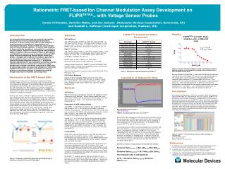
Ratiometric FRET-based Ion Channel Modulation Assay Development on FLIPRTETRA™ with Voltage Sensor Probes Carole Crittenden, Jennifer McKie, and Joe Jackson (Molecular Devices Corporation, Sunnyvale, CA) and Randall L. Hoffman (Invitrogen Corporation, Madison, WI) Results Figure 3. Barium chloride inhibition of inward rectifying potassium channel response in rat basophilic leukemia cells as determined by VSP ratiometric assay dyes. Barium chloride inhibited the Kir channel in rat basophilic leukemia cells at an IC50 concentration of 270 mM. This was calculated from the 4 parameter curve fit program in Graphpad Prism. Patch clamping data showed an IC50 concentration of 500 uM2. As a measure of the overall performance of the assay, Z’ was calculated at the IC80 value and equaled 0.58. Z’ factor is a tool for the comparison and evaluation of the quality of assays useful for assay optimization.3 A Z’ factor > 0.5 is considered a valid assay. An example of an assay data screen can be seen in figure 4. Conclusions A prototype configuration of 400 nm excitation LEDs and emission filters at 440-480 nm and 565-625 nm were used in FLIPRTETRA to effectively demonstrate ratiometric Voltage Sensor Probe assays. Ratiometric dye assays and FLIPRTETRA, along with other membrane potential reagentsare important tools for ion channel target assay development. A variety of user exchanged LED modules and filter combinations together with ratiometric dyes and simultaneous read 96-, 384- and 1536-well plate formats provides flexibility in measuring changes in membrane potential brought about by ion channels and transporters. Figure 4. FLIPRTETRA Screenworks data traces at 460 nm grouped by concentration. Lt. Green= Buffer Control Blue = Positive Control Apricot on RH side = No cells control FLIPRTETRA Instrument Setup Parameters Table 1. Ratiometric setup parameters for FLIPRTETRA Introduction Ion channels and transporters are proteins that regulate the movement of ions across biological membranes. Research on these proteins has led to the development of drug therapies for arrhythmias, hypertension, and neurological disorders. These proteins are also potential sites for adverse drug reactions. To screen large numbers of drug candidates for efficacy and safety, it is essential to use HTS assays. FLIPRTETRA was configured with prototype 400nm excitation LEDs and emission filters at 440-480 nm and 565-625 nm and was used to develop a 384-well FRET-based HTS assay employing Voltage Sensor Probe dyes, (VSPs). Changes were induced in membrane potential of rat basophilic leukemia cells, RBL-2H3, containing an inward rectifying potassium channel (Kir). Potassium chloride depolarized the cell membrane and barium chloride inhibited this response. Ratiometric analysis provided background correction and direct comparison of FRET data. In a prototype model, we show that utilization of VSPs with FLIPRTETRA provides a sensitive and efficient cell-based system for measuring changes in membrane potential brought about by ion channels and transporters. Mechanism of the FRET-based VSPs Voltage Sensor Probes are a Fluorescence Resonance Energy Transfer (FRET) based assay technology used for high-throughput ion channel drug discovery. The membrane-bound, coumarin-phospholipid (CC2-DMPE) FRET donor binds only to the exterior of the cell membrane. The FRET acceptor is a mobile, negatively charged, hydrophobic oxonol [either DiSBAC2(3) or DiSBAC4(3)], which binds to either side of the plasma membrane in response to changes in membrane potential. Resting cells have a relatively negative potential. Therefore, the two probes associate with the cell membrane exterior, resulting in efficient FRET. Exciting the CC2-DMPE donor probe (at ~405 nm) generates a strong red fluorescence signal (at ~580 nm) from the oxonol acceptor probe. When the membrane potential becomes more positive, as occurs with cell depolarization by KCl, the oxonol probe rapidly translocates (on a sub second time scale) to the other membrane face. Thus, each oxonol probe "senses" and responds to voltage changes in the cell. This translocation separates the FRET pair, and exciting the CC2-DMPE donor probe now generates a strong blue fluorescence signal (at ~460 nm) from the CC2-DMPE probe. Materials VSP Solution 1 160 mM NaCl (Cat# S 5886), 4.5 mM KCl (Cat# P 5405 ), 2 mM CaCl2(Cat# C 7902), 1 mM MgCl2(Cat# M 4880), 10 mM glucose (Cat# G7021) All from Sigma, St. Louis, MO, 10 mM HEPES (Cat# 15630-080, Invitrogen, Carlsbad, CA), pH 7.4 High K+ solution 164 mM KCl (Cat# P 5405 ), 2 mM CaCl2 (Cat# C 7902), 1 mM MgCl2(Cat# M 4880), 10 mM glucose (Cat# G7021) All from Sigma, St. Louis, MO, 10 mM HEPES (Cat# 15630-080, Invitrogen, Carlsbad, CA), pH 7.4 DMSO (Cat# 47,230-1 Aldrich, St. Louis, MO) Barium Chloride (Cat# B 0750, Sigma, St. Louis, MO) Voltage Sensor Probes Set: DisBAC2(3) and CC2-DMPE (Cat# K1016), VABSC-1 Background Suppression Dye (Cat# K1019 all from Invitrogen, Carlsbad, CA) RBL-2H3 Rat Basophilic Leukemia Cells (Cat# CRL-2256, ATCC, Manassas, VA) Cell Culture Reagents Eagle’s Minimum Essential Media (Cat# 30-2003, ATCC, Rockville, MD), 10% FBS (Cat# SH-30071, Hyclone, Logan, UT), Trypsin/EDTA (.25% Trypsin/1mM EDTA) (Cat# 25200-072), Dulbecco’s PBS (Cat# 14040-133, both from Gibco, Carlsbad, CA) Methods Cell Culture RBL-2H3 cells were trypsinized, washed, and plated at 12,500 cells/well in a BD BioCoat™ Poly-d-Lysine coated 384- well plate (Cat# 356663, BD Biosciences, Franklin Lakes, NJ) 18 to 24 hours prior to assay. A no cell control was placed in Column 24 A-F. Preparation of VSP loading buffers CC2-DMPE: A 5 mM stock solution of was prepared in DMSO and stored at –20o C. On the day of the assay, a working solution was prepared. An equal volume of 10% Pluronic acid was mixed with CC2-DMPE and subsequently diluted to 5 mM in VSP-1 solution. The buffer was vigorously mixed and protected from light prior to use. DisBAC2(3): A 12 mM stock solution was prepared in DMSO and stored at –20o C. A 200 mM stock solution of VABSC-1 was prepared in water and stored at RT. On the day of the assay, a 4 mM working solution of DisBAC2(3) was prepared including 0.21 mM VABSC-1 quencher in VSP-1 solution. No special considerations were necessary to utilize the VSP dyes on FLIPRTETRA. Loading Cells Media was removed from all wells of the 384 well plate and replaced with 50 ml VSP-1 solution. VSP-1 was removed immediately and replaced with 25 ml CC2-DMPE loading buffer and incubated at room temperature for 30 minutes, covered and protected from light. CC2-DMPE loading buffer was removed after 30 minutes and wells were washed with 50ml VSP-1 solution. VSP-1 was removed immediately and replaced with 4 mM DisBAC2(3) and 0.21 mM VABSC-1 loading buffer and incubated for 30 minutes at room temperature protected from light. A series of of BaCl2 (inhibitor) concentrations was added to respective wells at the start of the incubation period. On FLIPRTETRA,readings at both 460 nm and 580 nm were taken for 10 seconds before adding 25 ml High K+ solution to the wells and for about 40 seconds after addition. These form the basis of the Ratio. Instrument parameters are listed in Table 1. Calculation of Ratiometric Data Bias subtracted based on Sample 1 E460 nm= Red Color E580 nm= Blue Color Figure 2. Dual wavelength VSP Trace from FLIPRTETRA Ratiometric data analysis provides background correction and a direct comparison of FRET assay data at both 460 nm and 580 nm for each well. Data for the same cells can be compared in both polarized and non polarized states. An example of data from FLIPRTETRA is seen in Figure 2 above. A normalized ratio (Rf/Ro) reflecting background correction and RFU after addition of depolarizing agent divided by the initial RFU is calculated for each well. FLIPRTETRA is set to take readings at both 460 nm and 580 nm before and after addition of KCl buffer (depolarizing compound). Within identical data windows, an average of readings prior to addition and 40-60 seconds post addition are determined. Average readings from wells without cells are subtracted to calculate the baseline corrected values (BLC). Emission ratios for depolarized and polarized states: Emission RatioPolarized = BLC 460initial/BLC 580initial Emission RatioDepolarized = BLC 460final/BLC 580final The response ratio is calculated as: Rf/Ro = Emission RatioDepolarized/Emission RatioPolarized • References • M. Falconer et.al. High-Throughput Screening for Ion Channel Modulators, Journal of Biomolecular Screening, Volume 7, (5)460-465, (2002) • J. Gonzalez, et al.Cell-based assays and instrumentation for screening ion-channel targets, Drug Discovery Today, 4(9): 431-439, (1999) • J. Zhang, et. al. A Simple Statistical Parameter for Use in Evaluation and Validation of High Throughput Screening Assays, Journal of Biomolecular Screening, Vol. 4, (2)60-73, (1999) Figure 1. Schematic of VSP FRET-based dye indicating change in membrane potential (Courtesy of Invitrogen)


