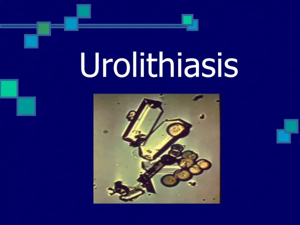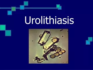
UROLITHIASIS
UROLITHIASIS. BACKGROUND. Urinary stone disease continues to occupy an important place in everyday urological practice. The average lifetime risk of stone formation has been reported in the range of 5-10%.

UROLITHIASIS
E N D
Presentation Transcript
BACKGROUND • Urinary stone disease continues to occupy an important place in everyday urological practice. The average lifetime risk of stone formation has been reported in the range of 5-10%. • A predominance of men over women (approx. 3:1) can be observed with an incidence peak between the fourth and fifth decade of life. • Recurrent stone formation is a common problem with all types of stones and therefore an important part of the medical care of patients with stone disease.
Theories of Stone Formation • A.Nucleation Theory • B.Stone Matrix Theory • C.Inhibitor of Crystallization Theory Most investigators acknowledge that these 3 theories describe the 3 basic factors influencing urinary stone formation. It is likely that more than one factor operates in causing stone disease. A generalized model of stone formation combining these 3 basic theories has been proposed.
RISK FACTORS •Start of disease early in life: <25 years •Stone containing brushite •Only one functioning kidney •Disease associated with stone formation: - hyperparathyroidism - renal tubular acidosis (partial/complete) - jejunoileal bypass - Crohn’s disease - intestinal resection - malabsorptive conditions - sarcoidosis - hyperthyroidism
RISK FACTORS •Medication associated with stone formation: - calcium supplements - vitamin D supplements - acetazolamide - ascorbic acid in megadoses ( > 4 g/day) - sulphonamides - triamterene - indinavir •Anatomical abnormalities associated with stone formation: - tubular ectasia (medullary sponge kidney) - pelvo-ureteral junction obstruction - calix diverticulum, calix cyst - ureteral stricture - vesico-ureteral reflux - horseshoe kidney - ureterocele
A). Disorders of urinary tract: congenital abnormalities those favor to apostasies; obstructive processes; neurogenic duskiness of the urinary tract; inflammative and parasitogenic damages; foreign bodies of urinary tract; traumatic injuries. B) Liver and digestive tract disorders: latent and manifested hepathopathiy; hepatogenic gastritis; colitis, etc. C) Endocrine diseases hyperparathyreoidism; hyperthyroidism; hypopituitaric diseases; D.) Infect focuses of the urogenital system. E) Metabolism disorders. essential hypercalciuria; disorders of membranes for colloid substances diffusion; renal rickets, etc F) Injuries those leads to continuous immobilization fractures of the vertebral column and limbs osteomyelitis diseases of the bones and joints chronic diseases of the visceral organs and nervous system. G) Climate and geographical causes. dry and hot climate with a high vaporization decrease water supply iodine deficiency H) Disorders of nutrition and vitamins balance: retinole and oscorbine acid deficiency in food. Excessive amount of the ergocalciferole in organism. Etiology (according Capital and I. Pogo Elko).
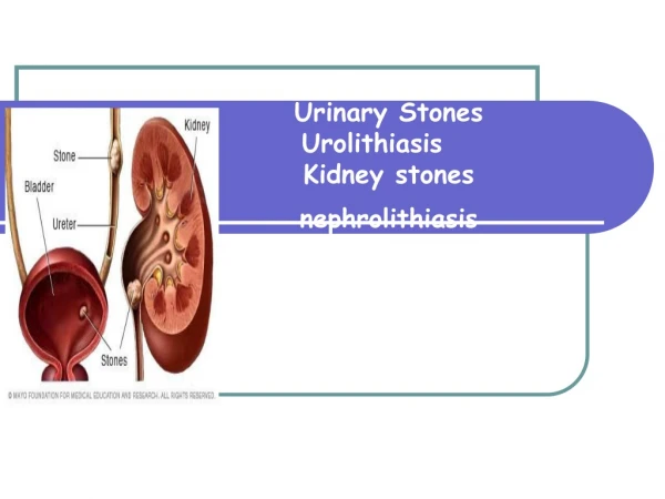
Renal Calculi • 1 Coral calculus • 2 Coral calculi fragment • 3 Calculi, which are impregnated with blood pigments
Medical History • A personal as well as a family history should be obtained for all patients. • A history of inflammatory bowel disease, recurrent urinary tract infection, prolonged periods of immobilization, gout, or familial occurrence of certain inherited renal diseases, eg, renal tubular acidosis or cystinuria, should be sought.
Clinical Manifestations • Acute obstruction of the urinary tract may cause renal colic, a form of severe abdominal pain often accompanied by nausea and vomiting due to celiac ganglion stimulation. Onset is sudden, often during the night or in the early morning
Clinical Manifestations • Obstructing calculi in the upper urinary tract cause an extreme crescendo like pain in the flank that generally radiates laterally around the abdomen to the corresponding groin and testicles in males and labia major in females. • When the stone obstructs the midureter, the pain tends to radiate to the lateral flank and abdominal region. • However, when the obstruction is in the distal ureter (near the ureterovesical junction), the patient exhibits symptoms of bladder irritation (frequency and urgency or genital pain).
Clinical Manifestations • Fever is rarely present except when a urinary tract infection accompanies obstruction. • Pulse rate and blood pressure, however, may be elevated as a result of the pain and agitation caused by the renal colic. • The patient's abdomen is generally flat and soft, with moderate deep tenderness on palpation where the calculus is lodged. • Some patients also have extensive hyperesthesia of the abdominal wall, either anteriorly or posteriorly. • The costo-vertebral area may be tender to percussion.
Laboratry Investigations • Stone analysis: In every patient one stone should • be analysed. • Blood analysis: Calcium Albumin Creatinine Urate • Urinalysis: Fasting morning spot urine sample • Dip-stick test: pH, Leucocytes/Bacteria • Cystine test, Ca, P, citrate, urate
Urinalysis. • This test usually reveals either gross or microscopic hematuria. Although hematuria may be absent in complete obstruction, microhematuria may be present in symptomatic partial obstruction. • Pyuria, usually moderate, may accompany obstruction even in the absence of identifiable infecting organisms. If severe pyuria is present, infection should be considered (especially in a female), since the stones may be secondary to infection.
Diagnostic imaging Routine examination involves a plain abdominal film of the kidneys, ureters and bladder (KUB) At least 90% of all renal stones are radiopaque and therefore readily visible on a plain film of the abdomen
Diagnostic imaging Excretory pyelography must not be carried out in the following patients - those: • With an allergy to contrast media • With S-creatinine level > 200 µmol/L • On medication with metformin • With myelomatosis
Diagnostic imaging Special examinations that can be carried out include: • Retrograde or antegrade pyelography • Retrograde pneumo-pyelography or cystography • Spiral (helical) unenhanced computed tomography (CT) • Scintigraphy.
Diagnostic imaging Ultrasonography- Inpatients in whom it is not possible to obtain an intravenous urogram, ultrasonic evaluation of the kidneys may aid in the diagnosis of renal stones. In pregnant women with flank pain in whom it is desirable to limit radiation exposure or in anuric patients or patients with chronic renal failure, the presence of hydronephrosis on acoustic shadowing may be diagnostic.
Diagnostic imaging • Cystoscopia shows swallowing of the ureter orifice in lower location of the stone, it may also partially project out to the orifice.
TREATMENT • Conservative • Instrumental • Surgical
Pain relief Pain relief involves the administration by various routes of the following agents: • Diclofenac sodium • Indomethacin • Hydromorphone hydrochloride + atropine sulphate • Baralgin • No-spae + Analgine • Tramadol
Pain relief • Warm bath • Spasmolytic “cocktails” (with papaverine, spasmalgone, no-spanum, promedole) should be taken. • A high dosage of the cystenal or urolesan (20 drops on the piece of sugar) is rather effective at the start of the renal colic. • If ache doesn’t disappear the novocaine blockade of the spermatic cord in males and round ligament in females is required. • Physical method.
Pain relief • For patients with ureteral stones that are expected to pass spontaneously, suppositories or tablets of diclofenac sodium, 50 mg administered twice daily over 3-10 days, might be useful in reducing ureteral oedema and the risk of recurrent pain. The patient should be instructed to sieve the urine in order to retrieve a concrement for analysis.
Pain relief • When pain relief cannot be obtained by medical means, drainage by stenting or percutaneous nephrostomy (PN) or stone removal should be carried out.
Stone removal The size, site and shape of the stone at the initial presentation influence the decision to remove the stone. Also, the likelihood of spontaneous passage has to be evaluated. Spontaneous stone passage can be expected in up to 80% of patients with stones not larger than 4 mm in diameter. For stones with a diameter exceeding 7 mm the chance of spontaneous passage is very low. The overall passage rate of ureteral stones is: • Proximal ureteral stones: 25% • Mid-ureteral stones: 45% • Distal ureteral stones: 70%
Indications for Active Stone removal Stone removal is usually indicated for stones with a diameter exceeding 6-7 mm. Active stone removal is strongly recommended in patients fulfilling the following criteria: • Persistent pain despite adequate medication • Persistent obstruction with risk of impaired renal function • Stone with urinary tract infection • Risk of pyonephrosis or urosepsis • Bilateral obstruction. • Obstructing calculus in a solitary functioning kidney
Stone removal • A test for bacteriuria should be carried out in all patients in whom stone removal is planned. Screening with dipsticks might be sufficient in uncomplicated cases. In others, urine culture is necessary. In all patients with a positive test for bacteriuria, with a positive urine culture or when there is suspicion of an infective component, treatment with antibiotics should be started before the stone-removing procedure. • Bleeding disorders and anticoagulation treatment should be considered. These patients should be referred to an internist for appropriate therapeutic measures during the stone-removing procedure. Treatment with salicylates should be stopped 10 days before the planned stone removal.
Indications to surgical operation • Frequent attacks of the renal colic or persistent pain that disables the patient. • Disorder of the urine outflow causing the hydronephrotic degeneration of the kidney. • Obturative anuria. • Frequent attacks of the acute pyelonephritis, progress of the chronic pyelonephritis that causes renal insufficiency. • Total hematuria. • Calculous pyonephrosis, apostematous pyelonephritis or carbuncle of the kidney. • Stone at the sole kidney that causes obstruction. • Stone in the ureter of the sole kidney that won’t pass away spontaneously.
Stone removal • In patients with coagulation disorders the following treatments are contra-indicated: extracorporeal shock wave lithotripsy (ESWL), percutaneous nephrolithotomy with or without lithotripsy (PNL), ureteroscopy (URS) and open surgery. • In pregnant women, ESWL, PNL and URS are contra-indicated. In expert hands URS has been successfully used to remove ureteral stones during pregnancy, but it must be emphasized that complications of this procedure might be difficult to manage. • In such women, the preferred treatment is drainage, either with a percutanous nephrostomy catheter, a double-J stent or a ureteral catheter . • For patients with a pacemaker it is wise to consult a cardiologist before undertaking an ESWL treatment.
Percutaneous Procedures • Percutaneous nephrostomy. Because of this technique, urologists can now perform operative procedures within the kidney without using the standard large flank incisions and mobilization of the kidney. • This technique, along with refinements in endoscopic instruments and advances in fiberoptics, allows endoscopic manipulation in the upper urinary tract by the percutaneous approach. • Percutaneous nephrolithotomy with or without lithotripsy (PNL)
Closed Surgical Procedures • Cystoscopic technique [With the patient under anesthesia and with fluoroscopic control, stones in the distal ureter can sometimes be removed with a wire stone basket] • Ureteropyeloscopy [Manipulation of small ureteral stones under direct vision with a ureteroscope is a major advance in the management of ureteral calculi. With this technique, small stones can be easily trapped in a stone basket and safely extracted through the dilated ureter.
Extracorporeal Shock Wave Lithotripsy • An extracorporeal noninvasive technique that uses shock waves to disintegrate urinary calculi while the patient is immersed in a water bath has been tested extensively and is now in clinical use. With this technique, calculi in the upper urinary tract are reduced to fragments, which pass spontaneously from the collecting system and bladder in most patients. • Size, location, and consistency of stone determine the number of shocks needed for fragmentation. In general, between 500 and 2,000 shocks arc necessary to fragment and pulverize an intrarenal calculus sufficiently for complete passage.
Open Surgical Procedures • Pyelolithotomy:Simple pyelolithotomy is used for removal of calculi confined to the renal pelvis. Minimal dissection of the renal sinus is usually needed, and exposure of the entire kidney is not required. This procedure is not indicated for the removal of entrapped caliceal stones or large, branched renal calculi.
Open Surgical Procedures • Ureterolithotomy. There are retroperitoneal, transperitoneal and combined surgical accesses. It depends on stone location. To remove stone from the superior ureter the Fedorov’s access is used, from medial ureter – Cuckulidze’s or Derev’yanko access is performed, the inferior ureter – Pyrogov’s access is needed, the pelvic portion of ureter may be accessed through the suprapubic arcuate incision.
Open Surgical Procedures NephrectomyNephrolithotomyCystolithotomy
Preventive treatment in calcium stone disease Preventive treatment in patients with calcium stone disease should be started with conservative measures. Pharmacological treatment should be instituted only when the conservative regimen fails. Patients should be encouraged to have a high fluid intake. This advice is valid irrespective of stone composition. For a normal adult, the 24-h urine volume should exceed 2000 ml, but the supersaturation level should be used as a guide to the necessary degree of urine dilution. The fluid intake should be evenly distributed over the 24-h period, and particular attention should be paid to situations where an unusual loss of fluid occurs.
Preventive treatment in calcium stone disease Diet should be of a 'common sense' type - a mixed balanced diet with contributions from all food groups but without excesses of any kind. The intake of fruits and vegetables should be encouraged because of the beneficial effects of fibre. Care must be taken, however, to avoid fruits and vegetables that are rich in oxalate. Wheat bran is rich in oxalate and should be avoided. In order to avoid an oxalate load, the excessive intake of products rich in oxalate should be limited or avoided. This is of particular importance in patients in whom high excretion of oxalate has been demonstrated. The following products have a high content of oxalate : • Rhubarb 530 mg oxalate/100 g • Spinach 570 mg oxalate/100 g • Cocoa 625 mg oxalate/100 g • Tea leaves 375-1450 mg oxalate/100 g • Nuts 200-600 mg oxalate/100 g.
Preventive treatment in calcium stone disease Vitamin C in doses up to 4 g/day can be taken without increasing the risk of stone formation. Animal protein should not be ingested in excessive amounts. It is recommended that the animal protein intake is limited to approximately 150 g/day. Calcium intake should not be restricted unless there are very strong reasons for such advice. The minimum daily requirement for calcium is 800 mg and the general recommendation is 1000 mg/day. Supplements of calcium are not recommended except in cases of enteric hyperoxaluria, in which additional calcium should be ingested with meals.
Preventive treatment in calcium stone disease The intake of foodstuffs particularly rich in urate should be restricted in patients with hyperuricosuric calcium oxalate stone disease , as well as in patients with uric acid stone disease. The intake of urate should not be more than 500 mg/day. Below are examples of food rich in urate : • Calf thymus 900 mg urate/100 g • Liver 260-360 mg urate/100 g • Kidneys 210-255 mg urate/100 g • Poultry skin 300 mg urate/100 g • Herring with skin, sardines, anchovies, sprats 260-500 mg urate/100 g.