Download
1 / 24
240 likes | 269 Views
Learn about the digestive system, its functions, key organs, and the process of digestion and absorption in the alimentary canal. Explore the structure and function of the mouth, teeth, salivary glands, pharynx, esophagus, stomach, and small intestines.

E N D
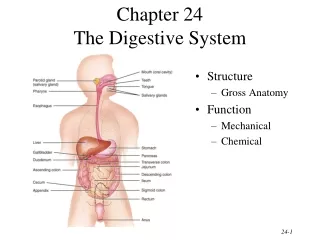
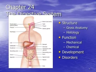
Chapter 15 - The Digestive System • Irregular tube; open at both ends, called “Alimentary canal” or “Gastrointestinal (GI) Tract” • 29 feet long (adults) - 9 meters • Food & other substances that enter tube are not really inside body • Passageway of food: broken down (digested) and absorbed thru walls < entering body - cells • Both - Mechanical & Chemical Digestion
Break Down of Food • Teeth- first physical breakdown • Stomach-churning of food (physical) • Mouth- first chemical breakdown (salvia) • Digestive enzymes throughout GI tract • Digestion - Process where large food particles reduced to absorbable molecules • Absorption - Process of small molecules passing thru digestive system walls into body
Key Organs of the GI Tract • Know Main & Accessory organs, Table 16-2; page 476 • Small Intestine : Duodenum, Jejunum, & Ileum • Large Intestines (elimination > feces): • Cecum, Colon: Ascending, Transverse, Descending, Sigmoid
Wall of Digestive Tract - • Mouth to anus • Four layers of tissue; surrounding the hollow space within the tube “lumen” • May vary in structure in different organs • Mucosa or mucous membrane - tough in esophagus, delicate, for absorption or secretion in rest of tract • Submucosa - connective tissue, blood vessels & nerves
Muscularis - 2 layers, responsible for wavelike, rhythmic contractions (peristalsis), moves contents, assists in mixing & mechanical breakdown • Serosa - outermost covering, composed of visceral peritoneum • Mesentery - double folded peritoneal tissue, anchors loops of digestive tract to posterior wall of abdominal cavity
Mouth - • Oral cavity - hollow chamber (roof, a floor, & walls) • Entrance of food; digestion begins immediately • Mucous membranes > mucus, protects against digestive juices & lubricates food passage • This mucous protects & lubricates • Hard palate - bony structure, front portion • Soft palate - posterior, chiefly muscles • Uvula - cone-shaped process hanging down from soft palate. W/ help of soft pal., prevents food or liquid from entering nasal cavity
Floor of the mouth - • Tongue - skeletal muscular structure, covered w/ mucous membrane • Anchored to bones in skull > hyoid bone • Frenulum- thin membrane; attaches tongue to floor of mouth • Tongue-tied: too short • Papillae: small elevations on surface • Vallate type - largest, inverted V-shaped row of about 10-12 mushroomlike elevations - tastebuds
Teeth - • Four major types - • Incisors - (sharp/cutting) • Canines - cuspids (pierce/tear) • Premolars - bicuspids & Molars - tricuspids (grinding/crush) • Mastication > chewing of food • Forms a bolus > ready for swallowing • By age 2 - full set 20 teeth (cut 1st - 2 yrs.) • By age 17 to 24 - 32 permanent teeth (cut 1st - 6 yrs.)
Typical Tooth - • Three main parts - • Crown - visible, covered w/ enamel (hardest tissue in body) • Neck - narrow portion surrounds by gum tissue (gingiva) • Root - fitted into socket in upper or lower jaw, lined by fibrous, periodontal membrane • Inside Structure - Enamel on outside, Dentin, Pulp cavity (blood vessels & nerves) moving inward
Salivary Glands - • 3 Pairs - ducts drain saliva into oral cavity, secretes about 1 liter/day • Parotid - in front of each ear (mumps - tender) • Submandibular - ducts by fernullum • Sublingual - ducts into floor of mouth • Saliva contents - salivary amylase (begins CHO digestion), mucus (moistens food)
Pharynx - • Behind nasal cavity & mouth • Tubelike structure made of muscles, lined w/ mucous membrane • Part of respiratory & digestive systems • Esophagus - • Passage for food to stomach • Tube-like structure, 10 inches long • Mucous lined • GERD - often caused by hiatal hernia
Stomach - • Upper part of abdominal cavity, under diaphragm • Pouch for food, hollow, expands (can push up on diaphragm > discomfort) • Lower esophageal sphincter (LES) or cardiac sphincter - rings of muscle tissue at end of esophagus - keeps food from reentering the esophagus when the stomach contracts
Chyme - semi-solid mixture, produced by contraction of stomach muscles that mixes food w/ gastric juices • Stomach contractions - • Created by 3 layers of muscles, run lengthwise, around, obliquely • Makes stomach one of strongest organs > peristalsis • Breaks food into tiny particles
Mucous membranes line stomach - contains gastric glands > secrete gastric juice & hydrochloric acid • When empty, wrinkled folds - rugae • Three divisions of stomach - • Fundus, body, pylorus • Pyloric sphincter - holds food in stomach, empties contents slowly into small intestine
Ulcer - carterlike wound or sore in membrane of stomach • 1 in 10 persons suffer in USA • Helicobacter pylori bacterium (H. pylori) • Small Intestines - • Portion of digestion tract that extends from the pylorus to the ileocecal valve • 12- 20 feet in length, coiled, convoluted, and occupies most of the abdominal cavity • Intestinal glands - secrete digestive juices • Smooth muscle wall - contracts > peristalsis
Plicea - circular folds covered w/ villi, increases surface area > absorption • In or on the villi - • Blood capillaries - absorb CHO & protein end products (glucose & amino acids) • Lacteal - lymphatic vessel - absorb lipids • Microvilli - brushlike border, > surface more • Chemical digestion - most occurs in 1st subdivision of duodenum • Minor & major duodenal papillae - ducts where pancreatic enzymes & bile enter small intestine
Liver - • Large organ, fills R upper abdominal cavity • Exocrine gland - secretes bile into ducts • Hepatic - means liver • Bile - essential for breaking up or emulsification of fats • CCK (cholecystokinin) - hormone secretion triggered by lipids in chyme > makes gallbladder contract & release bile • Drains from common bile duct into duodenum • Gallbladder - concentrates & stores bile
Pancreas - • C-shaped, exocrine gland that lies behind the stomach & duodenum • Pancreatic juice - most important digestive juice - contains enzymes for all 3 food groups • Sodium bicarbonate (alkaline substance) - neutralizes hydrochloric acid • Enters small intestine thru same duct as bile • Islets of Langerhans - hormones produced • Pancreatitis - inflammation (blockage, CF)
Large Intestine - • Begins with the ending of the ileum at the ileocecal valve - called the cecum • Approximately 5 feet in length, much larger in diameter than small intestine • Contents - not called chyme • Function - reabsorb water & salts • Material acted on by bacteria > more nutrients from cellulose & other fibers • Synthesis Vit. K needed for blood clotting, • Production of some B-complex vit.
Not as well suited for absorption as small intestine - no villi • Normal passage of material thru large intestine - 3 to 5 days • Subdivisions - flow in GI Tract one-way • Cecum - pouchlike area • Ascending colon - right side of body • Bends at hepatic or right colic flexure • Transverse colon - extends across front • Bends at splenic or left colic flexure • Descending colon - left side abdomen
Sigmond colon - S-shaped segment, terminates in rectum • Anal canal - terminal end of rectum, ends at external opening - anus • Inner anal sphincter - involuntary, smooth muscle, keeps anus closed except during defecation • Outer anal sphincter - striated, voluntary muscle
Appendix - • Vermiform appendix - “worm-shaped”, tubular structure, blind tube • No important digestive fnc. - digest cellulose • Appendicitis - inflammation • Peritoneum - • Large, moist, slippery sheet of serous membrane • Peritoneal space - small space between parietal & visceral layers - surfaces slide freely • Retroperitoneal - organs outside peritoneum • Extensions of peritoneum-mesentary, greater omentum - both assist in anchoring abd. contents
Digestion -Chemical & mechanical breakdown • CHO - amylase in mouth, slight effect • amylase from pancreas - into small intestine • Absorption of simple sugars (glucose) • Proteins - stomach (HCL/pepsinogen> pepsin) • Finished in small intestine by pancreatic (trypsin) & peptidases in intestinal juice • Amino acids - basic protein units
Fats - in small intestine • Bile emulsification of fats > pancreatic lipase > fatty acids & glycerol • Key digestive juices & enzymes • * page 410 Table 15-2 • Absorption - taking food, breaking it down in form for utilization of body • Just as important as digestion