Download
1 / 35
350 likes | 371 Views
Learn about Saudi Lung Cancer Prevention and Screening Guidelines to combat the high incidence of lung cancer in Saudi Arabia. These guidelines cover primary prevention, early detection, and screening recommendations to reduce the burden of this disease.

E N D
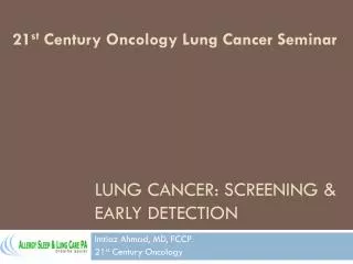
Saudi Lung Cancer Prevention and Screening Guidelines On behalf of Lung Cancer Prevention Guidelines Subcommittee Prof. Abdul Rahman Jazieh, MD, MPH Chairman, Department of Oncology King Abdulaziz Medical City Ministry of National Guard Health Affairs
INTRODUCTION Lung Cancer is number one cancer killer worldwide afflicting the lives of 1.8 million annually. In Saudi Arabia, in 2014, Lung cancer ranked fourth among Saudi males and seventeenth among Saudi females. There were 452 documented cases of lung cancer accounted to 3.9% of all newly diagnosed cases among Saudis in 2014. Lung cancer affected 354 (78.3%) males and 98 (21.7%) females with a male to female ratio of 361:100. The ASR was 5.3/100,000 for males and 1.4/100,000 for females In the United States the incidence of lung cancer (estimated 234,030; 54.6 per 100,000, data for 2011-2015)
I- Primary Lung Cancer Prevention II- Early Detection of Lung Cancer III- Lung Cancer Screening Guidelines Sections
I- Primary Lung Cancer Prevention II- Early Detection of Lung Cancer III- Lung Cancer Screening Guidelines Sections
I- Primary Lung Cancer Prevention The prevention of lung cancer is centered around tobacco control. This daunting task is the responsibility of multiple entities from national governing bodies all the way down to the individual level. These guidelines focus on the health care professionals’ role in prevention.
Management Guidelines for Tobacco Control for Health Care Providers Stop smoking cessation interventions and services should be available in primary care and community settings for everyone over the age of 12. The aim of these guidelines is to ensure that everyone who smokes is screened for smoking and is given the required advice and motivation to stop and all the assistance they might need for quitting and preventing relapse
1. General Measures Use local statistics to estimate smoking prevalence among the local population Prioritize specific groups who are at high risk of tobacco-related harm. Assure that your facility is smoke free facility and your patient and staff are not smoking in areas that are not designated for smoking. If you are a smoker, stop smoking, seek help if needed. It is advisable for your own, your family’s, and your patients’ health. You will be a good role model to your patients. It was found that smoking physicians are less likely to counsel their patients about smokingAcquire and provide smoking cessations tools and materials in your facility. Attend smoking cessations training workshop, if possible. Identify local/community resources to refer your patients, when needed.
2.Screening patients for smoking Ask all your patients systematically if they smoke or not. Make it part of their vital signs. Monitor stop smoking services by setting targets, including the number of people using the service and the proportion who successfully quit smoking. If a smoker is identified, implement smoking cessation guidelines of point 3
Smoking Cessation Intervention Table 2. the ‘’5 A’s” model for treating tobacco use and dependence If a patient is identified as a current smoker, counseling should be provided Provide smoking cessation counseling using the 5A technique (Ask, Advise, Assess, Assist, and Arrange) or any other proven approach. (Table 2)
Table 3. Enhancing motivation to quit tobacco, the “5 R’s” If the patient is not ready to quit, use motivational intervention. To motivate the patient consider quit attempt with the “5 R’s” (Relevance, Risk, Reward, Roadblocks, and Repetition). (Table 3)
Cognitive Behavioral Therapy and self-management technique should be used. Enlist the help of available resources in your community. For adults use Varenicline, Bupropion or nicotine replacement therapy (NRT). Agree a quit date set within the first 2 weeks of Bupropion treatment and within 1-2 weeks of Varenicline treatment. Agree a quit date if NRT is prescribed. Ensure that the person has NRT ready to start the day before the quit date. Ensure follow-up and behavioral support is provided. Ensure at least a very brief advice is delivered. Monitor compliance by auditing patients’ self -reported abstinence records, using objective measures such as carbon monoxide monitoring. Success is defined as less than 10 parts per million (ppm) at 4 weeks after the quit date. This should not imply that treatment should stop at 4 weeks.
I- Primary Lung Cancer Prevention II- Early Detection of Lung Cancer III- Lung Cancer Screening Guidelines Sections
II. Lung Cancer Early Detection Early detection of lung cancer means to discover the cancer as early as possible by recognition of the possible early signs and symptoms of cancer and doing the proper work-up or referral to confirm or rule out cancer. Early detection may occur proactively by screening asymptomatic patients for cancer. This section will focus on patients who present with symptom, signs, or findings suggestive of lung cancer, which should trigger “early diagnosis” of the disease.
All patients If a patient is asymptomatic and older than 55 with 30 PY history of smoking, follow Guidelines on section III. Keep in mind that non-smokers and females can get lung cancer too.
Findings that are suspicious suggestive of lung cancer Symptoms: new onset of New cough lasting more than 3-4 weeks, or a change in the “smoker’s chronic cough” Hemoptysis: especially not accompanying Upper Respiratory Infection (URI) or bronchitis, symptoms > 4 weeks Chest pain Shortness of breath Poor appetite/Weight loss
Signs of physical examination findings include: Cachexia Respiratory distress Clubbing Facial and neck edema and neck venous distension Signs of pleural effusion Signs of mediastinal shift Supraclavicular or cervical adenopathy
Imaging findings in asymptomatic and symptomatic patients (incidental findings): Mass with or without cavitation Pleural effusion Atelectasis/volume loss Mediastinal deviation or widening Lytic bone lesions Normal X-ray Keep in mind that chest x-ray can be normal in patients with lung cancer. Therefore, if a patient had symptoms such as hemoptysis > 4 weeks and chest x-ray is normal, obtain CT of chest and even if the CT is normal, patient may need bronchoscopy to diagnose an endobronchial small lesion. Recurrent pneumonia in the same lobe.
4. Laboratory findings include: • Hypercalcemia • Hyponatremia
3. Work-Up 3.1. Obtain good quality chest X-ray to rule out other causes of symptoms such as pneumonia or Tuberculosis. 3.2. Obtain CT scan of the chest for the following indications • Abnormal chest x-ray, suspicious of cancer • Persistent infiltrate despite antibiotic treatment for a pneumonia • Normal chest x-ray with concerning complaints such as persistent hemoptysis or cough > 4 weeks 3.3. Thoracentesis: For a pleural effusion in the setting of suspected malignancy. 3.4. Bronchoscopy • If normal CT scan with suspicion of endobronchial lesions. • If there is accessible lesion. • Include endobronchial ultrasound staging for hilar and mediastinal nodes if possible in the absence of distant metastasis 3.5. CT guided biopsy : For peripheral nodules or masses not accessible by bronchoscopy
I- Primary Lung Cancer Prevention II- Early Detection of Lung Cancer III- Lung Cancer Screening Guidelines Sections
III. Lung Cancer Screening Lung cancer screening is proven to save lives and became the standard of care in the United States especially after National Lung Cancer Screening Trial (NLST). However, screening studies were done among a Western population where lung cancer is very prevalent and kills more people than breast, prostate and colon cancer combined. But given the lower prevalence in our population such screening cannot be automatically presumed to be successful or cost effective to be adopted. However, since we are practicing most of our medicine, based on published evidence, we propose here stringent guidelines to provide to our health care professionals who might like to take care of their patients based on available evidence in the literature and consider providing them with lung cancer screening. It is very prudent that health care professional be aware of all the requirements (Table 4) and challenges of doing screening program before launching screening to all his patients.
1. Requirements for lung cancer screening program: These requirements should be considered prior to starting screening patients (Table 4). 1.1. Who should be screened (Section 2) 1.2. Where will CT scan of chest done, would the radiologist be willing to do it, who will pay for it, will the report be standardized, who responsible to do follow-up plan based on the report? 1.3. What would ordering physician do if there was abnormal CT scan findings. Do they have multidisciplinary team with expertise to manage suspicious findings or have access to tertiary center that one can refer the patient to them for further work-up. Confirm with the center if they accept all your referrals and how many and who is eligible for referral and the referral process? (See Figure 1 as prototype) 1.4. Who would call the patient and report the result and follow-up on the patient work-up? 1.5. Who would counsel the patient for medical and psychological issues? 1.6. Must have access to/or offer smoking cessation counseling to smokers? 1.7. If diagnosed with cancer, where will the patient be treated? 1.8. If there was incidental finding such as coronary artery disease or emphysema, where would physician refer patient?
Table 4: Requirements for local lung cancer screening program or individual requests
2. Whom to screen: The following are candidates for lung cancer screening: 2.1. Asymptomatic patients (symptomatic patients should be worked up properly according to standards of care) 2.2. Age 55 – 77 years 2.3. Smoking history > 30 PY (number of packs smoked per day x year of smoking) 2.4. Active smoker or quit smoking less than 15 years ago. 2.5. Did not have chest CT scan the last year 2.6. Do not perform screening for individuals with comorbidities that could adversely influence their ability to tolerate the evaluation of screen-detected findings or tolerate treatment of an early-stage screen-detected lung cancer, or that substantially limit their life expectancy. We recommend that low-dose CT screening should not be performed in these situations. (Strong recommendation, low-quality evidence) (e.g. advanced liver disease, COPD with hypoventilation and hypoxia, NYHA class IV heart failure)14
3. What to do? 3.1. Explain to patient the screening process and potential risks and benefits and follow up plans in case the patient opts for screening. 3.2. Obtain low dose CT scan (LDCT) of the chest 3.2.1. CT examinations should be acquired using multidetector scanners with at least 16 detector rows, in a helical technique 3.2.2. Intravenous contrast material should NOT be used. 3.2.3. Field of view: From the lung apices to the costophrenic angles. 3.2.4. Section thickness should be 2.5 mm or less, with less than 1 mm preferred 3.2.5. Gantry rotation should be 500 msec per rotation or faster. 3.2.6. Dose: Maximum dose threshold as a volumetric CT dose index of 3 mGy for a standard-sized patient (height [170 cm]; weight 70 kg) with “appropriate dose reduction” for smaller patients and an appropriate increase for larger patients 3.2.7. Optimum Low dose protocol are established separately depending on vendor specific parameters and adjustments to match dose threshold of CT dose Index of 3 mGy
3.3. Lung-Rad lung imaging reporting and data system is to be used in reporting as established by American College of Radiology 3.4. Based on the report, decide next step (Section 4 and 5) 3.5. A positive test is defined as a test that leads to a recommendation for any additional testing other than to return for the annual screening exam. 14
4. Managing findings on the screening CT scan (for primary care/non-specialist) 12 14 17 4.1. Normal CT scan: after 1 year and then annually repeat CT scan 4.2. If CT scan is positive per 3.4, compare with a previous imaging, if available. 4.3. New lung lesions < 5 mm repeat in 12 months. Repeat annually if no increase in size 4.4. New lesions 6-7 mm repeat CT scan in 6 months 4.5. New lesions > 8mm refer to pulmonary/thoracic surgeon for work-up 4.6. Other findings 4.6.1. Coronary artery disease (CAD) refer to cardiology 4.6.2. Chronic obstructive pulmonary disease (COPD) – assess need to refer to pulmonary 4.6.3. Other finding: act based on findings
5. Work-up of lung cancer nodules (to be done by Consultant with access to multidisciplinary team) 5.1. A multidisciplinary team including at least one consultants from each of the following specialties: radiology/interventional radiology, pulmonary/interventional pulmonary and thoracic surgeon. Team should review all the cases and take consensus approach to decision making. 5.2. New solid lesion > 8 mm obtain CT Scan with IV contrast or PET/CT scan 5.2.1. PET positive or suspicious CT scan obtain CT guided or surgical excisional biopsy (resection). 5.2.2. PET negative or low suspicion, repeat LDCT in 3 months, if still stable, repeat LDCT in 6 months, if still stable, do LDCT scan annually. If it grew, biopsy or surgical resection.
5.3. Management of part-solid lung nodules 5.3.1. <5 mm: annual LDCT 5.3.2. >6mm with <5 mm solid component: LDCT in 6 months 5.3.3. >6mm with 6-7 mm solid component: LDCT in 3 months or PET scan 5.3.4. > 8 mm solid component: CT chest with IV contrast , if highly suspicious: biopsy or surgical excision 16
5.4 New non-solid Lesion: 5.4.1 < 19 mm repeat LDCT in 6 months, if stable annual LDCT, if grew biopsy or surgical resection 5.4.2 > 20 mm repeat LDCT in 3 months, if stable repeat in 6 months then annual. If grew, biopsy of surgical resection.
5.5 Management of multiple lung nodules: 5.5.1 Purely solid: measure and manage the largest and as solid nodules 5.5.2 Nodules with partly solid component: measure and manage the largest as part solid nodule