Download
1 / 40
470 likes | 718 Views
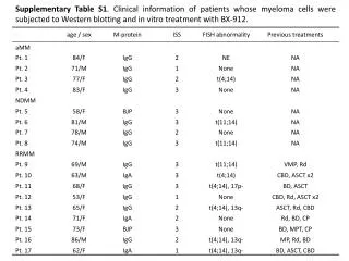
Understand the role of imaging in multiple myeloma patient management, including precise bone disease identification for treatment initiation, differential diagnosis, and follow-up. Learn the advantages of PET/CT in identifying small lesions, extra-medullary disease, and treatment evaluation.

E N D
Evidence for use of CT scanning and PET/CT in managing patients with myeloma Elena Zamagni “Seragnoli” Institute of Hematology Bologna University School of Medicine
ROLE OF IMAGING IN MULTIPLE MYELOMA • Precise identification of bone disease, as sign of organ damage and need to start treatment • Identification of sites of extra-medullary disease (total body techniques) • Differential diagnosis between localized disease (BSP) and systemic disease (MM) • Correct identification of sites of bone disease at risk of complications (fractures, neurological complications) (MRI gold standard) • Correct follow up of the patients after treatment, in particular in non secretory MM Zamagni E. et al, BJH 2012
ACTIVE MYELOMA: the CRAB CRITERIA • C:Calcium levels increased • R:Renal insufficiency • A:Anemia • B: Bone lesions, osteolytic or osteoporosis Myeloma-related end organdamage due to the plasma cell proliferative process IMWG, BJH 2003 Rajkumar V. et al., Lancet Oncology 2014
INTERNATIONAL MYELOMA WORKING GROUP UPDATED CRITERIA FOR THE DIAGNOSIS OF MULTIPLE MYELOMA • Definition of myeloma bone disease (CRAB):clearevidence of one or more sites of osteolytic bone destruction (atleast 5 mm or more in size) seen on CT, WBLDCT, PET/CT, regardless of weatherthey can be visualized on skeletalradiography or not • Ifdoubtlesions on CT or PET/CT: close follow-up every 3-6 months and/or biopsy of the lesion • Oseoporosis per se in the absence of lyticlesionsisnotsufficientfor CRAB Rajkumar V. et al., Lancet Oncology 2014
IMAGING TECHNIQUES IN MM: WBXR Whole body X Rayhasalwaysbeen the standard for the evaluation of bone disease, however: Lyticlesions are visibleonlyifatleast 30%-50% of trabecularsubstanceislost Unable to identify small osteolyticlesions (planar technique) Lowsensitivity in the spine Unable to distinguishbetweenosteoporoticvertebralfractures and MM relatedones Itcannot be used for the assessment of response to treatment Frequent underestimation of MM bone disease Zamagni E. et al, BJH 2012 Pianko et al, Clin Canc Res 2014
ROLE OF NEWER IMAGING TECHNIQUES • MORPHOLOGICAL: assessing bone destruction • WB-MDCT-LDCT, CT part of PET/CT • FUNCTIONAL: assessing bone marrow infiltration and disease metabolism • ASSIAL MRI- WBMRI (DCE-MRI, DWI-MRI), PET/CT • Active MM • at diagnosis: staging and prognosis • after treatment: evaluation of treatment response • Early stage/smoldering MM
WHOLE BODY LOW-DOSE MULTIDETECTOR ROW-CT (WB-LDCT) • Fast scanning time, low radiation dose (3,3-7 msV), high resolution images • Demonstration of extra-osseus findings Shortt CP et al, Sem Musculoskel Radiology 2010 Ippolito D. et al, Eur J Radiol 2013 Wolf MB et al, Eur Journal Radiology 2014 Pianko MJ et al, Clin Canc Res 2014 Horger M., EJ Radiol, 2004 Hur J., J Comput Assist Tomogr, 2007
WBLDCT vs WBXR IN THE STAGING OF MM Pianko MJ et al. Clin Cancer Res 2014
ADVANTAGES OF THE USE OF PET/CT IN MM • Capability to identify small osteolytic lesions (usual resolution limit: 5 mm) • Possibility to detect both medullary and extra-medullary disease and to localize it with anatomic precision by the use of CT • Capability to distinguish between active or inactive disease and/or necrosis-fibrotic tissue • Capability to identify infections or other tumors
EXTRAMEDULLARY DISEASE (EMD) • Need to identify true EMD from para-medullary/breakout lesions • Incidence ranging from 7% to 18%; more frequent in later phases of the disease 1,2 • Increased incidence in the last years due to the availability of more sensitive imaging techniques and the prolongation of survival 1,2,3 • Extremely poor prognosis even in the novel agents era 1,2,3,4,5 • Associated with unfavorable cytogenetic abnormalities and GEP defined high-risk MM 5 • Well assessed by PET/CT and whole body techniques (WB-DWI MRI); in a recent meta-analysis higher sensitivity and diagnostic accuracy of PET/CT for EMD 4 1 Varettoni M. et al, Annals of Oncology 2010 2 Bladè J. et al, JCO 2011 3 Wale A et al, Haematologica 2016 4 Lu Y.Y. et al, Clinical Nuclear Med 2012 5 Usmani S.Z. et al, Haematologica 2012
COMPARISON OF PET OR PET/CT AND CONVENTIONAL IMAGING AT STAGING • 18 studies, 798 patients • 7 studies PET CT vs WBXR: 6/7 PET showed more lytic lesions with the exception of the skull • 5 studies PET CT vs MRI spine and/or pelvis: 4/5 MRI was superior in detecting myeloma bone disease, especially in case of diffuse bone infiltration • 1 study PET/CT vs WBMRI: concordant in 80% cases • Identification of extra-medullary disease Van Lammeren-Venema D et al., Cancer 2011
32 directly comparison studies, prospective and retrospective, 1661 patients • Index test vs reference standard: detection rate • Quality assessment of diagnostic studies • All index tests had sensitivity above 0,9 as compared to WBXR (low false negative). Fewer additional lesions detected by PET/CT and MRI as compared to WBLDCT WBLDCT can replace WBXR • Modern imaging techniques detected fewer lesions in the skull and ribs «We therefore recommend additional X-ray of the ribs and the skull if clinically relevant» Regelink J. et al., BJH 2013
PROGNOSTIC RELEVANCE OF PET/CT AT DIAGNOSIS (EFS AND OS) IN ASCT CANDIDATES 1,2,3 • Correlation between imaging techniques and standard prognostic factors in univariate and multivariate analyses; the strongest was PET/CT FLs and beta2-mic, LDH and CRP1,2 • Correlation between PET/CT FLs and GEP derived variables, such as high-risk designation (70-gene model) and PR subtypes 1,2 • PET/CT FLs, LDH and cytogenetics abnormalities retained independent prognostic value for PFS and OS in Cox regression analysis 1 Bartel. TB et al, Blood 2009 3 Usmani S.Z. et al, Blood 2013 2 Waheed S et al, Haematologica 2012
PFS PFS 1.00 1.00 1.00 1.00 64% at 4 yrs 60% at 4 yrs PFS 43% at 4 yrs 0.80 0.80 0.80 25% at 4 yrs 0.80 P= 0.0008 P= 0.008 65% at 4 yrs 0.60 0.60 0.60 0.60 43% at 4 yrs N° FLs ≤ 3 OS N° FLs > 3 P= 0.01 89% at 4 yrs 0.40 0.40 0.40 0.40 65% at 4 yrs SUV ≤ 4.2 SUV > 4.2 Extramedullary neg Extramedullary neg Extramedullary pos Extramedullary pos P= 0.006 0.20 0.20 0.20 0.20 0.00 0.00 0.00 0.00 0 12 24 36 48 60 72 84 0 12 24 36 48 60 72 84 0 0 12 12 24 24 36 36 48 48 60 60 72 72 84 84 months months months months PROGNOSTIC VALUE OF PET/CT AT DIAGNOSIS IN ASCT CANDIDATES N° OF FLs, SUV VALUE, EMD SUV value N° FLs EMD EMD Zamagni E. et al, Blood 2011
PROGNOSTIC VALUE OF PET/CT AT STAGING Resultsreproduced in: • Several independent series of patients ASCT candidates, correlating with MRI findings, standard prognostic factors and molecular features of PCs1,2,3,4 • Small group of patients non ASCT eligible (retrospective study) 5 • Series of patients pre- ALLO SCT (retrospective study) 6 • Re-staging at relapse (retrospective studies) 7,8 1 Zamagni E. et al, Blood 2011 5 Zamagni E. et al, Clin Canc Res 2015 2 Bartel. TB et al, Blood 2009 6 Patriarca F. et al, Biol BMT 2015 3 Waheed S et al, Haematologica 2012 4 Usmani S.Z. et al, Blood 2013 7 Lapa C. et al, Oncotarget 2014 8 Derlin T. et al, EJNM Mol Imag 2011
IMAGING TECHNIQUES AT DIAGNOSIS IN ACTIVE MM: WBLDCT PROS CONS • Sensitivity and specificity • 3D structural info for CT-guided biopsy, surgery, RT planning • Can depictEMD, BM involvement, lytic lesions • Can assess tumor burden • Rapid acquisition time, low radiation dose • Comfortable for patients • Inexpensivecompared to PET/CT or MRI • Lesions in ribs and skull may be missed • Sub-optimal for diffuse bone marrow involvement • Few data/unclear prognostic significance of lesion number* • More expensive compared to WBXR • Radiation exposure > WBXR *Nishida Y et al., Blood Canc J 2015
IMAGING TECHNIQUES AT DIAGNOSIS IN ACTIVE MM: PET/CT PROS CONS • Sensitivity and specificity • Optimal to assess EMD • Can depict lytic lesions (CT part) • Can assess tumor burden and disease metabolism • Prognostic significance of FLs and SUV • Useful for staging of SPB • Sub-optimal for diffuse bone marrow involvement and skull lesions • Cost> WBLDCT, WBXR and MRI • Radiation dose > WBXR, WBLDCT • Availability
IMAGING TECHNIQUES AT DIAGNOSIS • In both every-day practice and clinical trials WBLDCT is suggested to substitute soon WBXR to define bone end- organ damage • WBXR may be used if WBLDCT is not avaliable • Always consider WBLDCT or PET/CT when WBXR is negative and there is bony pain • PET/CT is a valuable tool for the detection of osteolyses and metabolic lesions predicitng outcomes. Outweight these benefits with economic sustainability • In patients without lytic lesions and CRAB criteria, MRI (at least axial) is recommended for the possible detection of FLs (new diagnostic criteria) Zamagni E. et al, BJH 2012 Regelink JC et al, BJH 2013 Terpos E et al, Haematologica 2015 Pianko MJ et al, Clin Canc Res 2014 Mesguich C et al, EJR 2014 Dimopoulos M et al, JCO 2015
ROLE OF FDG PET/CT IN MULTIPLE MYELOMA • Symptomatic MM • at diagnosis: staging and prognosis • after treatment: evaluation of treatment response, follow-up • Early stage/smoldering MM
METABOLIC RESPONSE TO THERAPYPROGNOSTIC VALUE OF PET/CT BEFORE ASCT Bartel. TB et al, Blood 2009 • Complete FDG suppression retained independent prognostic value for PFS and OS in Cox regression analysis Usmani S.Z. et al, Blood 2013
METABOLIC RESPONSE TO THERAPY PET-CT normalisation following 3 cycles of RVD Impact on PFS (32% normalised) 78.7% 54.8% p = 0.04 Moreau P. et al, ASH 2015
1.00 1.00 PFS 47% at 4 yrs 0.80 0.80 32% at 4 yrs P= 0.02 months 0.60 0.60 OS SUV 100% reduction 79% at 4 yrs 0.40 SUV < 100% reduction 0.40 66% at 4 yrs P= 0.02 0.20 0.20 months 0.00 0.00 0 12 24 36 48 60 72 84 96 108 120 0 12 24 36 48 60 72 84 96 108 120 METABOLIC RESPONSE TO THERAPYPROGNOSTIC VALUE OF PET/CT AFTER ASCT MULTIVARIATE ANALYSIS Zamagni E. et al, Blood 2011
METABOLIC RESPONSE TO THERAPYPET/CT MRD MONITORING IN CR PATIENTS ASCT eligible and not-eligible (189 pts) ASCT candidates (192 pts) Zamagni E. et al, Blood 2011 • 70% PET-CR, 40-50% biochemical CR • 25-30% of the patients in conventionally-defined CR had PET/CT still positive Zamagni E. et al, Clin Canc Res 2015
METABOLIC RESPONSE TO THERAPY PET-CT normalization before maintenance Impact on PFS (62% normalised) 69% 51,6% p < 0.001 Moreau P. et al, ASH 2015
METABOLIC RESPONSE TO THERAPY PET-CT normalization before maintenance Impact on OS (62% normalised) 94,6% 69,9% p = 0.003 Moreau P. et al, ASH 2015
METABOLIC RESPONSE TO THERAPY PET-CT normalization before maintenance PFS in Arm A: RVD x 8 cycles PFS in Arm B: frontline ASCT Adjusted on otherprognosticfactors p = 0.009 Univariate log-rank, p = 0.027 Adjusted on otherprognosticfactors p = 0.01 Univariate log-rank, p = 0.01 Adjusted on otherprognosticfactors p = 0.008 Univariate log-rank, p < 0.001 OS in Arm B: frontline ASCT Moreau P. et al, ASH 2015
METABOLIC RESPONSE TO THERAPY PET/CT and MFC MRD MONITORING BEFORE MAINTENANCE 89,6% PFS PET/CT and MFC neg 54,5% • 86/134 evaluated by both PET/CT and flow • 47,7% both negative PET/CT and/or MFC pos p = 0.02 Moreau P. et al, ASH 2015
PROGNOSTIC VALUE OF PET/CT AFTER TREATMENT Caldarella C. et al, Int J Mol Imaging 2012
IMAGING TECHNIQUES AFTER TREATMENT: PET/CT PROS CONS • Specificity • Earlier post-therapy changes • Prognostic significance in CR patients (MRD monitoring) • Good correlation with biochemical response • Lack of standardization • Applicability in 75% of the patients • Availability, cost Zamagni E. et al, BJH 2012 Hillengass J. et al, Leuk and Lymphoma 2013 Mesguich C et al, EJR 2014
IMAGING TECHNIQUES AFTER TREATMENT OPEN ISSUES • Do we need the same imaging technique at baseline and after treatment to evaluate metabolic response? • Is the persistence of «severe» positive imaging findings during treatment allowing to change therapy and do we need to evaluate it? • Should we tailor treatment (consolidation/maintenance) on imaging-defined minimal residual disease? • Which relationship between bone marrow MRD and imaging MRD?
FOLLOW-UP PHASE AFTER FIRST-LINE TREATMENT Usefullness of PET/CT follow-up • 282 pts, studiedat baseline and after treatment (novel agents +/- ASCT) with PET/CT (every 12-18 months) • Median follow-up after first-line treatment: 56 months • 63% of the patients with relapse/progression: • 37% onlyserologic • 48% serologic + skeletal • 15% onlyskeletal • 88% clinical (pain, pathologicalfractures) and PET/CT • 12% exclusive PET/CT (7% of the pts, 11% of allprogressions) Zamagni E. et al, Clin Canc Res 2015
60 54 48 42 36 30 24 18 12 6 0 Pet suv 4.2-6 Pet suv <4.2 Pet suv6+ PROGNOSTIC VALUE OF PET/CT AFTER TREATMENT CORRELATION BETWEEN TTP AND SUVmax post ASCT months Correlation coefficient = -0,67, P= 0,008 Cuzick’s trend test P= 0,017 Nanni C. et al, Clin Nuclear Medicine 2012 Zamagni E. et al, Clin Canc Res 2015
NEWER IMAGING TECHNIQUES: FOLLOW-UP POST TREATMENT • Serial evaluation with novel imaging techniques (PET/CT or WBMRI) after first line treatment is currently not recommended to all the patients because of the high costs (monetary and radiation exposure) and low usefulness. Our analysis confirm this suggestion • In the small subgroup of patients with a persistent high glucose metabolism after first-line treatment (approximately 10%), serial PET/CT evaluation can be recommended during the follow-up, in order to point-out possible progression not otherwise identifiable
ROLE OF FDG PET/CT IN MULTIPLE MYELOMA • Symptomatic MM • at diagnosis: staging and prognosis • after treatment: evaluation of treatment response, follow-up • Early stage/smoldering MM
TTP TTP PET/CT neg (97 pts): 30% at 2 yrs PET/CT neg: median 60 mos PET/CT pos (74 pts): 75% at 2 yrs PET/CT pos with osteolyses (16 pts): median 21 mos (87% at 2 yrs) p = 0.0008 p = 0.004 Siontis B. et al, Blood Cancer J 2015
Time to progression of SMM to active MM 100 75 PET/CT negative 50 PET/CT positive 25 HR 3.00 (95% CI 1.58-5.69) p=0.001 0 0 1 2 3 4 Years PROGNOSTIC ROLE OF FLs WITHOUT OSTEOLYSES IN SMM Median 4,5 yrs Median 1,1 yr Prospective study on 120 pts, median f up 2,2 years Centralized imaging revision 16% pts with FLs, without underlying osteolytic lesions Probability of progression at 2 years PET/CT pos pts vs neg: 58% vs 33% Zamagni E. et al., Leukemia 2015
EXTENSIVE USE OF NEWER IMAGING TECHNIQUES OPEN ISSUES • Quality of many studies hampered by a poor description of selection and execution criteria • Major inconsistency in methodology between studies • Need to define standardized criteria for imaging definitions and positivity cut-off • IMWG prospectivestudy on WBLDCT • Italian/Europeanproposal for PET/CT interpretative criteria (Nanni C et al, EJNM 2015) Zamagni E. et al, BJH 2012 Regelink JC et al, BJH 2013 Pianko MJ et al, Clin Canc Res 2014 Mesguich C et al, EJR 2014
A visual degree of uptake is defined for the target lesion and extramedullary lesions according to the scheme proposed in Deauville Criteria for the evaluation of lymphoma patients Description, in a5 points scale, of: • Bone Marrow metabolic state (BM) • Number and site of Focal Lesions (Fx)with or without Osteolytic Lesions (Lx) • Presence and site of Extramedullary Disease (EM) • Presence of Paramedullary Disease (PM) • Presence of Fractures (Fr) Nanni C. et al., EJNM 2015
CONCLUSION • Newer imaging techniques have proved reliable tools in the staging and as predictors of outcome in MM patients, both in early stage and active disease • At diagnosis, WBLDCT is suggested to substitute in the near future WBXR • PET/CT and DWI-MRI are the favorite techniques for assessing and monitoring response to therapy and are becoming complementary investigation tools for detecting minimal residual disease, going beyond the conventionally defined CR level • Implementation of prospective clinical trials with newer imaging techniques will help to adress several issues, standardize the interpretation of the results and optimize the use of these promising tools. This may improve disease management