Hypothermia / Hyperthermia / Exposure
480 likes | 1.16k Views
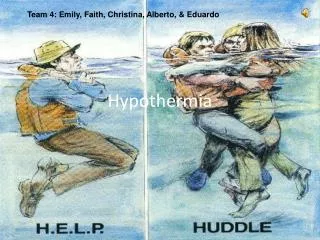
Hypothermia / Hyperthermia / Exposure. JP SMILES. Hypothermia. Core temp < 35 ° C Research limited to either mild hypothermia in healthy subjects or case reports. Pathophysiology. Heat loss occurs through Radiation Conduction Convection Evaporation

Hypothermia / Hyperthermia / Exposure
E N D
Presentation Transcript
Hypothermia / Hyperthermia / Exposure JP SMILES
Hypothermia • Core temp < 35 °C • Research limited to either mild hypothermia in healthy subjects or case reports
Pathophysiology • Heat loss occurs through • Radiation • Conduction • Convection • Evaporation • Hypothermia results in derangement of multiple organ systems • Shivering – increases metabolic rate but only while glycogen stores last and down to temps of 30 °C
CVS Effects • Initial tachycardia and peripheral vasoconstriction • Subsequent bradycardia (refractory to atropine), hypotension and fall in cardiac output • Osborn J waves appear < 32 °C • Anti-arrythmic drugs and inotropes/vasopressors are generally ineffective at temperatures < 30 °C
CNS Effects • Loss of fine motors skills and co-ordination then loss of gross motor skills • Progressive decrease in GCS • Cerebrovascular auto regulation is lost at 24 °C • 20 °C EEG is flat and patient appears dead as cerebral metabolism falls • Temperatures at which shivering is lost varies widely 24 °C - 35 °C • Temp < 28 °C = rigidity, mydriasis, and areflexia
Resp Effects • Initially rise in resp rate followed by depression and basal metabolic rate slows • CO2 retention and resp acidosis can occur • Significant fall in O2 consumption and CO2 production (50% at 30 °C) • Apnoea can develop • Initial left shift of the oxygen dissociation curve • Impaired O2 delivery and tissue hypoxia • Lactic acidosis • If acidosis becomes severe the curve shifts back R again
Renal Effects • Cold induced diuresis • GFR falls as CO and renal blood flow fall • ARF in 40% of patients who require ICU • Initial hypokalaemia due to shift of extracellular potassium into cells • Hyperkalaemia can occur with acidosis secondary to cell death
GI Effects • Intestinal motility decreases below 34 °C • Ileus < 28 °C • Oral medication is not appropriate • Hepatic impairment can occur due to reduced CO (Raised lactate and therefore Hartmans is a bad idea) • Pancreatitis and Mesenteric Venous Thrombosis are both common
Haem Effects • Increased blood viscosity fibrinogen and haematocrit • Coagulopathy may develop
Grading of Hypothermia • Mild (35 °C - 32 °C) • Moderate (32 °C - 28 °C) • Severe (<28 °C) • Temperature measurement • Accurate low reading digital of mercury thermometer • Placed 15 cm rectally of oesophageally (better as cold faeces can effect rectal temperatures)
Investigations • UEC • Hypo or hyperkalaemia/ARF/low HCO3- • Glucose • Hypo/Hyperglycaemia • CK • May be elevated • FBC • Increased haematocrit due to cold induced diuresis and hypovolaemia • Thrombocytopaenia • COAG • Coagulopathy and DIC is common • LFT • Transaminitis • LIPASE • Pancreatitis • VBG • Initial respiratory alkalosis • Secondary respiratory and metabolic acidosis
Investigations • ECG • Bradycardia • PR/QRS/QT prolongation • Variable ST and T wave changes • Osborn J waves • Arrythmias • AF/VT/VF/1st, 2nd, 3rd Degree HB
Osborn waves • These waves were definitively described in 1953 by JJ Osborn • Also called J waves • Delayed depolarisation • Represented as ST elevation at the QRS – ST junction • < 32 °C • Proportional to the degree of hypothermia • Not pathognomonic • SAH/Cerebral injuries/Myocardial ischaemia
Management • ABC • Remove wet clothing and insulate • Gentle handling – rough handling and invasive procedures have historically been thought to increase risk of cardiac arrythmias • Now thought these risks have been overemphasised • Consider co-existent pathology
Management • Intubation as necessary • IV Access (drugs IV only. IM SC poor absorption) • Urinary catheter • NGT • Temperature and cardiac monitoring • Fluid resuscitation • Dehydration is often present • Warmed fluids • Dextrose is good • Avoid drugs until core temp 30 °C – ineffective and may accumulate until released
Management Rewarming – mild hypothermia • Endogenous rewarming • Exercise if possible • Passive external warming • Warm dry environment • Cover with warm blankets
Management Rewarming – moderate hypothermia • Active external rewarming • Warm blankets • Radiant heat source • Bair hugger • 2°C per hour
Management Rewarming – severe hypothermia • Includes cardiopulmonary arrest • Warmed humidified inhaled oxygen • Warmed IV fluids • Warmed left pleural lavage • Warmed Peritoneal lavage • Cardiopulmonary bypass • Most other methods are ineffective
Management Arrythmias • VF may occur spontaneously in < 29 °C • Sinus brady and AF with slow ventricular response are common and can be considered physiological with hypothermia • AF usually reverts spontaneously on rewarming • Drugs and electricity are unlikely to work until temp is > 30 °C
Modification of ACLS for hypothermia • ETT – Warmed humidified air 42 °C - 46 °C • Aggressive active core warming • Warmed saline/peritoneal lavage/pleural lavage/bypass • VF/VT – Single defibrillation appropriate and initial drug therapy. If no response defer further attempts or drug doses until core rises above 30 °C • PEA/Asystole– Again wait till core temp above 30°C (atropine not likely to be effective) • Many anecdotal reports of unexpected survival • Not dead till they are warm and dead!!!!
Heat Related Illness • Heat stroke • Heat exhaustion • Heat cramps • These may occur as a continuum Bhut Jolokia pepper
Heat stroke • Core body temp > 40 °C • Hot dry skin • CNS abnormalities (delirium/coma)
Heat stroke • Classical – Occurs due to exposure to a high environmental temperature • Exertional – Occurs in the setting of strenuous exercise
Pathophysiology • Oxidative phosphorylation stops at temperatures > 42 °C • Cell damage • Loss of thermoregulatory compensatory mechanisms • Hypoxia, increased metabolic demands, circulatory failure, coagulopathies and inflammatory response
CVS Effects • Tachyarrythmias and hypotension • Two types exist with exertional heat stroke • Hyperdynamic group – high cardiac output and tachycardia • Hypodynamic group – Low cardiac output, increase peripheral vascular resistance
Neurological Effects • Cardinal features of heat stroke • Delirium, lethargy, coma and seizures • Can be permanent (up to 33%)
Rhabdomyolysis • Injured cells leak phosphate and calcium • Hypercalcaemia and Hyperphosphataemia • Hypokalaemia is seen early • Secondary to heat induce hyperventilation leading to respiratory alkalosis • Sweat and renal losses • Hyperkalaemia is seen later • Potassium losses from damaged cells and renal failure • Hyperuricaemia develops secondary to the release of purines from injured muscle
Renal Effects • ARF in approx 30% • Direct thermal injury to kidneys • Pre-renal insult of volume depletion and renal hypoperfusion • Rhabdomyolysis
Haematological • Exertional heat stroke is associated with haemorrhagic complications • Petechial haemorrhages or eccyhmosis secondary to direct thermal injury or DIC
Immunological • Similar to sepsis • The actions of inflammatory mediators account for the multi organ dysfunction
Assessment • Consider in patients with altered mental state and exposure to heat • Classic triad of hyperthermia, neurological abnormalities and dry skin • Measure temp with rectal/oesophageal probe • Sweating can still be present • Hypotension and shock 25% • Hypovolaemia, peripheral vasodilatation and cardiac dysfunction • Sinus tachy • Hyperventilation – a universal finding in heat stroke
Investigations • UEC • Hypokalaemia • Hyperphosphataemia and hypercalcaemia • Hyperkalaemia and hypocalcaemia may be present if rhabdomyolysis has occurred • Renal impairment
Investigations • Urate – is frequently high and may play a role in the development of acute renal failure • Glucose – elevated in up to 70% • LFT • Almost always seen in exertional heat stroke (AST and LDH most commonly elevated) • CK – 10000 to 1000000 in rhabdomyolysis
Investigation • FBC – WCC as high as 30 -40,000 • Coag – routinely abnormal and DIC may occur • Acid Base: • Lactic acidosis • Compensatory respiratory alkalosis • Myoglobin – serum or urine myoglobin may be elevated
Investigation • ECG • Rhythm disturbances (sinus tachy, SVT + AF) • Conduction defects (RBBB and intraventricular conduction defects) • QT prolongation (most common secondary to low K+ , Ca 2+ and Mg 2+) • ST changes (secondary to myocardial ischaemia)
Investigations • CXR: • ARDS • Aspiration
Management of Heat Stroke • If prompt effective treatment not undertaken mortality approaches 80% • A – ETT if needed • Consider early • Avoid suxamaethonium
Management of Heat Stroke • B • Monitor Resp Rate and O2 sats • Look for evidence of aspiration if GCS decreased • Check for ARDS and ventilate as per lung injury protocol • C • May be a large fluid deficit • N saline is probably best (CSL – lactate and avoid K+ containing fluids) • Monitor heart rate, BP, CVP and urine output • Picco/Swan-Ganz pulmonary artery catheter may be indicated • Pressors may be needed but avoid adrenergic agents as they can impair heat dissipation by causing peripheral vasoconstriction (dopamine)
Management of Heat Stroke • D – Intubate if needed • E – Temperature should be measured by oesophageal or rectal probe
Cooling Methods • Mainstay of therapy and must be initiated from the onset • Use prehospital may be lifesaving • Initially remove patient from heat source and remove all clothing • Evaporative cooling – tepid water on the skin with fans • Ice water immersion – most effective method but practically difficult and cant use monitors/equipment and uncomfortable for the patient
Cooling Methods • Ice packs to axilla, groin and neck • Cooling blankets and wet towels • Peritoneal lavage and cardiopulmonary bypass can be considered in severe resistant cases • Shivering may occur in rapid cooling – this will increase oxygen consumption and heat production • Sedate • paralyse • Paracetamol and aspirin are ineffective and should not be used
Outcome • Mortality should be less than 10% with prompt treatment • Most recover without sequalae • Residual neurological defects are reported
Heat Exhaustion • Heat exhaustion – mild heat stroke • Same physiological process • Patients can still have the capacity to dissipate heat and the CNS is not impaired • Volume depletion is still a problem
Heat Cramps • Painful involuntary spasms of major muscles • Usually in heavily exercised muscle groups • Dehydration and salt loss also thought to plat a role • Rest rehydrate and replace salts