Download
1 / 19
190 likes | 245 Views
Explore Trypanosomes, parasitic creatures with complex life cycles, impacting humans and domestic animals through blood-dependent transmission. Learn about Trypanosomatidae stages, species, transmission, and pathogenesis.

E N D
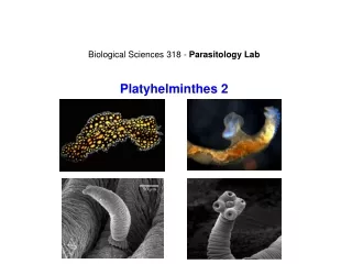
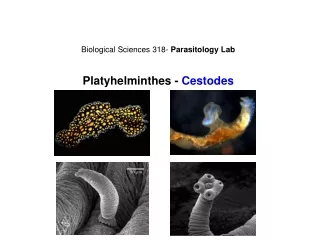
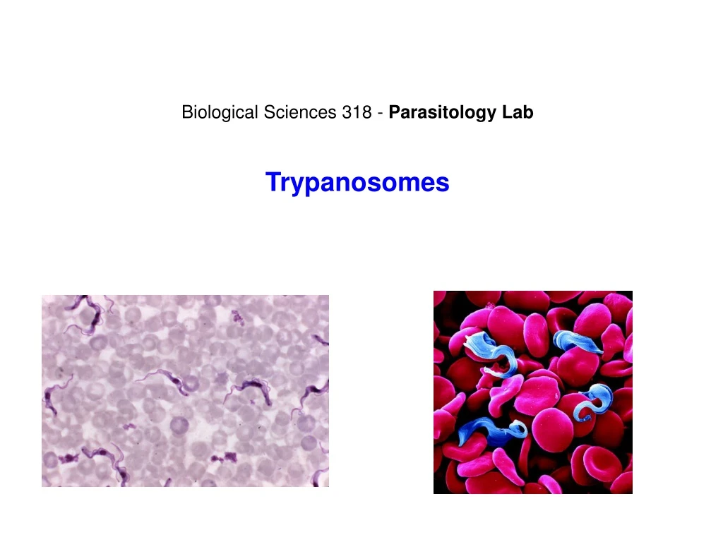
Biological Sciences 318 - Parasitology Lab Trypanosomes
Trypanosomes Kingdom II Euprotista Phylum Euglenista Order Kinetoplastida Three families: 1 - Bodonidae - free living 2 - Cryptobiidae - parasites of fish and invertebrates 3 - Trypanosomatidae - some members important to humans and domestic animals Heteroxenous (most) – require more than one host to complete life cycle Hemoflagellates – dependence on blood
Trypanosomatidae • Kinetoplast: • DNA containing organelle from which a mitochondrion arises; Mitochondrial compartment packed with minicircles and maxicircles of DNA; self-replicating. • 1 flagella • 1 nucleus • 1 mitochondrion • leaf-like or rounded body • all parastitic • change body form depending on host and tissue • cyclic development (6 different stages)
Trypanosomatidae Stages: Trypomastigote: Epimastigote: Promastigote: Amastigote: blood stream form; replicative stage infective stage of non-motile; infective form in insect Leishmania spp. intracellular, replicative stage in vertebrate
Genus Trypanosoma • some of the most economically important human and animal parasites • parasites of invertebrates and all classes of vertebrates • blood or tissue fluids; intracellular • mostly transmitted through invertebrate vectors • Development: • Anterior station - Salivaria • division of trypomastigotes in midgut of vector • migration of parasite forward into the upper digestive • tract (eg. salivary glands) • metacyclic trypomastigotes passed to vertebrate • host when the vector feeds. • Salivarian (eg.T.brucei gambiense) by tse-tse fly. • Posterior station - Stercoraria • parasite in the hindgut transforms into epimastigotes • and metacyclic trypomastigotes • move back through the digestive tract • metacyclic trypomastigotes are passed to the vertebrate • host in the vector feces. • Stercocarian (eg. Trypanosoma cruzi) by kissing bugs.
Trypanosoma brucei • Organism and Disease Associations, Host Range • Vector: Tse tse • Geographic Distribution and Importance Fly belt!
Trypanosoma brucei gambiense Lifecycle of Trypanosoma brucei gambiense: Anterior Station
Trypanosoma Slide:Trypanosoma brucei gambiense: blood smear Slide:Trypanosoma lewisi: parasite of rats. Ingestion of fleas or flea feces containing trypomastigotes Slide:Trypanosoma equiperdum: causes a venereal disease (dourine) in equines Transmission through sexual intercourse
Pathogenesis • General • Trypanosomes live in blood, lymph nodes, spleen – therefore not intracellular • Particularly abundant in intercellular spaces in brain • Humans • Local reaction: painful sore at site of bite, disappears after a couple of weeks • Trypanosomes reproduce rapidly once enter blood and lymph system – generalized invasion of all organs • Winterbottom’s sign – swollen nodes at base of skull • severe wasting • Somnolence • CNS signs – increased apathy, dullness, • tremors, convulsions, coma, death
Trypanosoma cruzi • Organism and Disease Associations, Host Range • = Schizotrypanumcruzi • Chagas disease, American Trypanosomiasis • Wild anddomesticanimals (reservoirs) • Vector: reduviidbugorkissingbug • Geographic Distribution andImportance Impact: 18-25 million cases Risk: 100 million in 21 countries 4th leading cause of mortality in Latin America 45,000 deaths per year directly attributed to Chagas leading cause of cardiac disease in S. and Central America
Trypanosoma cruzi Lifecycle of Trypanosoma cruzi: Posterior Station
Trypanosoma cruzi Slide:Trypansoma cruzi pseudocyst in rat cardia tissue. The pseudocyst is an enlarged cell containing amastigotes
Pathogenesis • General • Trypomastigote enter blood; • Invadehostcellsof RES, skeletalandcardiacmuscle • Cell breakdown, releaseofamastigotes; Infectionofadditonalcells • Productionofneurotoxinsaffectingconductingsystems • Muscleslooseabilitytocontract, Megacolon, - esophagus, heart
Genus Leishmania • Leishmania spp. Cause complex of infections displaying a variety of disease manifestations • Parasites of mammals, most commonly humans, dogs, rodents • Vector: Sandflies (Phlebotomus, Lutzomyia) • Organism and Disease Associations • Leishmania tropica (oriental sore or cutaneous leishmaniasis) • Leishmania donovani (Kala-azar or visceral leishmaniasis) • Leishmania braziliensis (espundia or mucocutaneous leishmaniasis) • Geographic Distribution and Importance • Currently the leishmaniases, prevalent in four continents, are considered to be endemic in 88 countries, 72 of which are developing countries: • 90% of all visceral leishmaniasis cases occur in Bangladesh, Brazil, India, Nepal and Sudan; • 90% of mucocutaneous leishmaniasis occurs in Bolivia, Brazil and Peru; • 90% of cutaneous leishmaniasis cases occur in Afghanistan, Brazil, Iran, Peru, Saudi Arabia and Syria.
Leishmania Lifecycle of Leishmania
Leishmania Slide:Leishmania donovani promastigote Slide: Leismania donovani in tissue smear: study the tissue smears (liver and spleen) containing amastigotes of L. donovani using oil immersion. The amastigotes are very small (2.5-5.0 μm) and spheroid to ovoid in shape. Only the nucleus and a very large kinetoplast are visible with light microscopy. Some describe amastigotes as little snowmen Slide:Leishmania tissue section replicates as intracellular amastigotes in macrophages. In fact, amastigotes will eventually kill a macrophage and go on to infect others.
Pathogenesis Visceral (L.donovani) This visceral disease has a new and old world form: particularly Brazil, and Mediterranean Europe, North Africa, East Africa, India and China. The amastigote forms are found within the reticulo-endothelial cells of the viscera, ie the spleen, lymph nodes, liver and intestine. The incubation time of 10 days-a year. The symptoms are a slow developing low grade fever, and general malaise, a progressive wasting of the patient with anemia. Other classic symptoms as the disease progresses is the protrusion of the abdomen, hepatospenomgegaly. If untreated death will occur within 2-3 years of contracting the infection. In acute forms, the disease can run its course within 6 - 12 months. Clinical signs include edema, particularly of the face, bleeding mucus membranes, breathing difficulties and diarrhea. Cutaneous (L.tropica) Usually localized to the site in which the sandfly bite occurs. The amastigotes multiply in the reticulo-endothelial system of the skin.
Leishmania Strategies of survival 1. When macrophages first engullf a parasite such as Leishmania, macrophages undergo a respiratory burst. This respiratory burst involves enzymes such as superoxides which should effectively digest the parasite. Some Leishmania sp can produce an enzyme called superoxide dismutase which can neutralize the free radical damage. 2. Leishmania has evolved the ability to bind Compliment-like C3 protein, which can outright inhibits the respiratory burst. 3. Macrophages function by communication usually via lymphokines, with T-cells. Leishmania are able to “shut down” communication between these cells by interrupting the transcription of major histocompatibility genes (MHC) so that the macrophages they are infecting bare few if any MHC proteins on the surface for T-cells to recognize. 4. Leishmania has been reported to stimulate the bone marrow to produce more macrophages. However, the new macrophages being produced are immature; they can capture and engulf the parasite but are physiologically unable to kill them, even in the presence of stimulating cytokines.
Learning Objectives • 1. Order Kinetoplastida • - Know general characteristics • 2. Family Trypanosomatidae • General characteristics (leaf-like, flagellum, parasites of vertebrates) • Undergo cyclic development as they pass from one host to the next • Know life cycle stages (purpose, morphology, differentiation) • 3. Genus Trypanosoma • General features • Anterior vs. posterior station • Visual ID T. lewisi, T. equiperdum, T. brucei gambiense, T. cruzi • 4. Trypanosoma lewisi • Host + vector transmission • 5. Trypanosoma equiperdum • Host + pathology + transmission • 6. Trypanosoma brucei gambiense • Distribution, pathology, vector • Life cycle (hosts, stages, transmission, tissue) • Vector + type of development in vector • 7. Trypanosoma cruzi • Pathology • Distribution, vector , life cycle, transmission • Visual ID pseudocyst in rat heart, what stage • 8. Genus Leishmania • Hosts, vectors, life cycle, transmission • Know 3 species of medical importance + disease associated with each • Visual ID promastigotes to genus for L. donovani • L. donovani amastigotes in liver tissue • What strategies does Leishmania use to overcome macrophage defenses Vocabulary Kinetoplast Amastigote Promastigote Trypomastigote Epimastigote Anterior station Posterior station Nagana Dourine Vector Reservoir host Definitve host Pseudocyst Macrophage Superoxides Respiratory burst Superoxide dismutase MHC