Download
1 / 36
360 likes | 488 Views
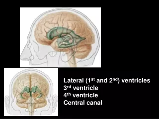
Lateral (1 st and 2 nd ) ventricles 3 rd ventricle 4 th ventricle Central canal. 4 th ventricle. Floor = rhomb oid fossa. Sulcus medianus Sulci limitantes fovea sup. (V) fovea inf. (IX, X) Trigonum n. XII

E N D
Lateral (1stand 2nd) ventricles 3rd ventricle 4th ventricle Central canal
4th ventricle Floor = rhomboid fossa Sulcus medianus Sulci limitantes fovea sup. (V) fovea inf. (IX, X) Trigonum n. XII Eminentia medialis colliculus facialis (VI) Striae medullares Recessus lat. (area vestibularis) Tuberculum acusticum sup. med. inf.
4th ventricle Roof = tegmen ■Velum medullare sup. ■Fastigium ■Velum medullare inf.,tela choroidea (pia mater+ependyma) + vessels = plexus choroideus Apertura mediana Aperturae lat.
Aqueductus mesencephali – 3rdventricle Walls: Rostral – columnae fornicis, commissura ant., lamina terminalis Superior - tela choroidea Posterior - recessus suprapinealis, com. habenularum, rec. pinealis, com. post.
Inferior – optic chiasma, infundibulum Lateral – thalamus, hypothalamus
Foramen interventriculare Rec. triangularis Rec.suprapinealis, pinealis Rec. opticus, infundibuli Aqueductus mesencephali
Pars centralis Cornu ant. (frontale) Cornu post. (occipitale) Cornu inf. (temporale)
corpus callosum septum pell. ncl. caudatus Cornu anterius
Cornu posterius calcar avis sulcus calcarinus em. collateralis sulcus collateralis
Cornu inferius: hippocampus eminentia collateralis corpus callosum tela choroidea
CT MRI
Meninges Calvaria Epidural space Ektomeninx (pachymeninx) -dura mater: periosteal, meningeal layers Subdural space arachnoid mater Endomeninx (leptomeninx) Subarachnoid space (CSF) pia mater
Dural infoldings: Falx cerebri Falx cerebelli Tentorium cereb. Diaphragma sellae Cavum trigeminale Vagina n. optici
Supply of the dura mater: Aa. meningeae Branches from: a. ethm. ant. – anterior cranial fossa a. maxillaris – middle cranial fossa a. phar. asc. – posterior cranial fossa CN V:supratentorial compartment C2, C3, CN X, XII:infratentorial compartment
Granulationes arachnoideales– protrude through the meningeal layer of the dura into the venous sinuses – transfer of CSF to the venous system
Pia mater Subarachnoid cisterns : • - chiasmatic • - fossae lat. cerebri • interpeduncular • ambient • quadrigeminal • pontocerebellar • - cerebellomedullary
Meninges of the spinal cord Epidural spaceDura mater spinalis Subdural spaceArachnoidea spinalis Subarachnoid spacePia mater spinalis: lig. denticulatum Pia mater Lig. dent. Arachnoidea Sp sub-a Dura mater Sp sub-d
Cisterna lumbalis Medullary cone: L1-2 Dural sac: S2-3 Lumbar puncture (spinal tap)
Liquor cerebrospinalis (CSF) Clear, colorless fluid,150 mL, secreted at the rate of 400-500 mL daily Produced by the choroid plexuses of ventricles Protects the brain, prevents the weight of the brain from compressing the nerves and vessels against the cranium. Circulation:Lateral ventricles – for. interventriculare – 3rd ventricle – aquaqeductus cerebri – 4th ventricle –median and lat. apertures – subarachnoid space –sinus durae matris
Arteries of the CNS Spinal cord Rr. spinales a. cervicalis asc. a. vertebralis a. cervicalis prof. aa. intercostales post. aa. lumbales a. iliolumbalis a. sacralis lat. a. sacralis mediana
Rr. spinales: aa. radiculares ant. et post.: a. spinalis ant. (fissura med. ant.) aa. spinales post. (sulcus lat. post.)
Vv. spinales Plx. venosi vertebr. int.> vv. intervertebrales >plx. venosi verteb. ext.> plx. suboccipitalis > vv. vertebrales vv. cervicales prof. vv. intercostales vv. lumbales vv. sacrales lat.
Brainstem, cerebellum Aa. cerebri post. Aa. cerebelli sup. Aa. pontis Aa. cerebelli inf. ant. A. basilaris Aa. cerebelli inf. post.Rr. ad med. oblong. Aa. spin. ant. et post. Aa. vertebrales
Brain Aa. cerebri: anterior (A. car. int.) media (A. car. int.) posterior (A. basil.)
a. cerebri ant. a. cerebri med. a. com.post. a. cerebri post. Circulus arteriosus ■ Aa. corticales ■ Aa. centrales■ Aa. choroideae
Veins of the brain ■ superficial Vv. cerebri sup. Vv. cerebri inf. V. cerebri media spf.
■deep veins 1. vv. septi pellucidi 2. vv. thalamostr. sup. 3. vv. choroideae sup. = Vv. cerebri int. + Vv. cer. med. prof.+ Vv. basales V. magna cerebri Sinus rectus
Sinus durae matris 2 3 1 rectus 2 sagittalis sup. 3 sagittalis inf. 4 occipitalis 5 transversus 6 sigmoideus 7 sphenoparietalis 8 petrosus sup. 9 petrosus inf. 10 cavernosus 11 plx. basilaris 6 1 5 4 7 10 9 11 8
Tributaries of sinuses Vv. ophtalmicae Vv. labyrinthi Vv. meningeae Vv. diploicae Vv. emissariae