Download
1 / 42
1.79k likes | 4.74k Views
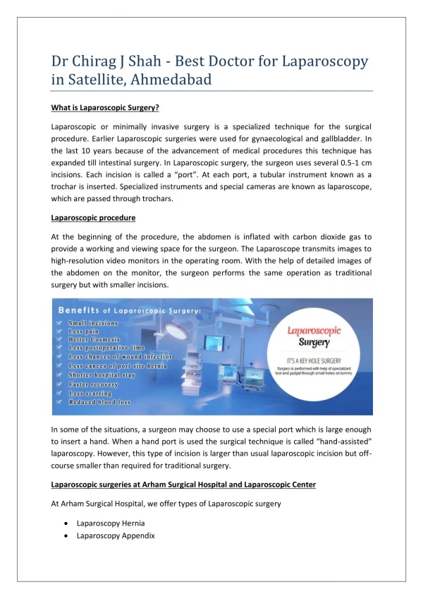
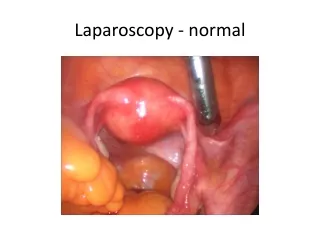
SAFE ENTRY IN LAPAROSCOPY. Yasser Orief M.D., PhD. Lecturer of Obstetrics & Gynecology, Alexandria University Fellow, L ϋ beck University, Germany DGOL, Auvergné University, France. Approximately 50% of all complications during laparoscopy are entry-related.

E N D
SAFE ENTRY IN LAPAROSCOPY • Yasser Orief M.D., PhD. • Lecturer of Obstetrics & Gynecology, Alexandria University • Fellow, Lϋbeck University, Germany • DGOL, Auvergné University, France
Approximately 50% of all complications during laparoscopy are entry-related. • One in 4 is not diagnosed during the operation Stoval&Mann,Up to date,May2011. Margina,Clin Obstet Gynecol2002,45:469 Seven top French centers 29 966 cases
Entry-Related complications Blood Vessel Injuries Small: Omental or mesenteric Major: Abdominal or pelvic artery or vein Epigastric vessel perforation Retroperitoneal hematoma due to vena cava injury with the Veress needle
Bowel Injuries Perforation of the small bowel by 10-mm trocar
Insufflation of the subcutaneous or preperitoneal space: 1-Subcutaneous emphysema. 2-Complicating visibility.
How to diminish entry-related complications?Surgeon’sexperience is the most important 1-Understanding of relvant anatomy. 2-Proper use of surgical instruments. 3-Choice of optimal surgical technique. 4-Ability to detect and manage complications Jansen et al. Complications of laparoscopy: a prospective multicentre observational study. Br J Obstet Gynecol. 2008
The Surgeon Should Follow The Safe abdominal entry guidelines for: • Position of the patient. • Placement of the veress needle. • Pneumoperitoneum. • Primary (umbilical) trocar insertion. • Accessory (secondary) trocars. • Identification of high risk patients.
High Risk Patients For Abdominal Entry Body weight(Obesity,slimmness). Large pelvic mass. Previous abdominal and pelvic operations. Strong abdominal musculature. Previous radiation therapy. Bowel distension.
Position of the Patient • The patient is positioned in the dorso-lithotomy position with lowered legs.
Position of the Patient • The operating table should be in the flat position • Early Trendelenburgposition may lead to major vascular injury.
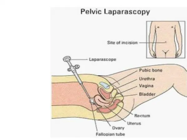
Placement of Veress Needle • The umbilicus is the ideal site of choice for insertion: • The thinnest area • Minimal subcutaneous fat even in obese women. • Fusion of fascial layers with the peritoneum
Placement of Veress Needle • Before insertion, the needle is checked for patency and spring mechanism • The angle of insertion is usually 90o to the skin, then it is readjusted according to thickness of abdominal wall: 45o in thin women to 90o in obese women.
Weight < 75 Kg Angle of isertion = 45°
95 kg > Weight > 75 Kg Angle of insertion = 45°- 90°
Weight > 95 Kg Angle of insertion = 90°
Veress needle Safety Tests Or Checks • Provide very little useful information on the placement of the veress needle. • It is therefore not necessary to be performed. SOGC Practice Guideline. 193, 2007
What Is The Most Reliable Safety Test ? • The Veressintraperitoneal(VIP) pressure ≤ 10 mm Hg • Therefore, it is appropriate to attach the CO2 source to the Veress needle on entry. SOGC Practice Guideline.193, 2007
Guidelines for Safe Veress Needle Abdominal Entry • Aim to sacral hollow. • Aim at right angle to the skin, then readjust. • Aim away from pelvic vessels. • Test for peritoneal entry (IPP ≤ 10 mm Hg). • Advance only 2-3 cm after piercing peritoneum. • Avoid over-insufflation. RCOG Guideline No 49 May 2008
Avoid: • Long Needle. • Premature Trendelnburg. • Distension: stomach, colon or bladder. • Adhesions.
Pneumoperitoneum • Adjust intra-peritoneal pressure to 12-16 mm Hg once insertion of trocar is complete. SOGC Practice Guideline. 193, 2007
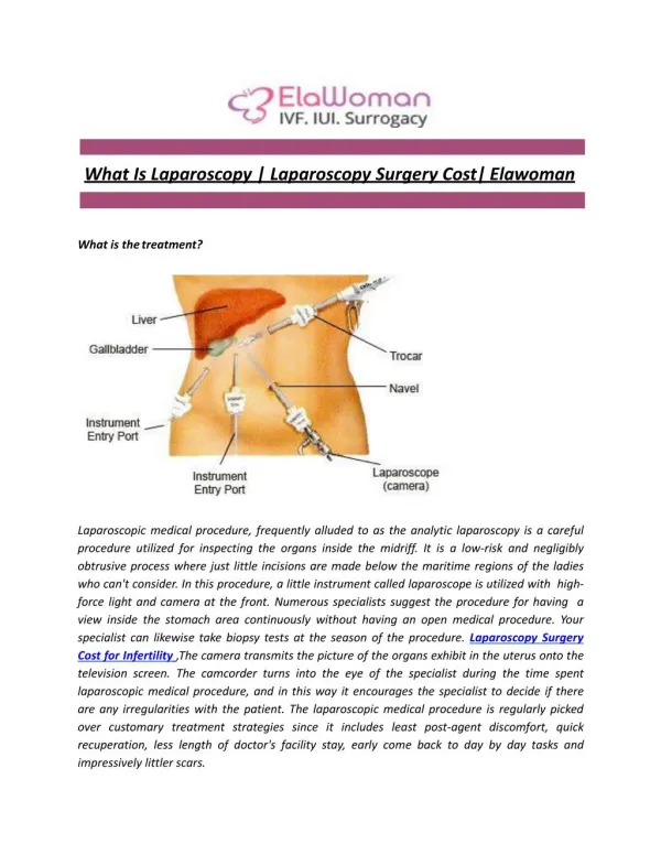
Types Of Primary Trocar 1-Non-Visual Entry System: • Conventional trocar and cannula • Disposable shielded trocars • Reusable shielded safety trocars • Radially expanding(Versa step) 2-Visual Entry Systems 3-Open laparoscopy (Hasson)
Trocar introduction The primary incision should be vertical from the base of the umbilicus RCOG Guideline no 49, May 2008
Guidelines for Safe Primary Trocar Entry • Aim at right angle to the skin, then readjust 45-90o. • Aim to sacral hollow,aim away from pelvic vessels. • Extend index on the trocar to prevent sudden thrust of trocar. • Rotate in a semi-circular fashion. • Advance not more than 2-3cm beyond peritoneum. RCOGGuideline No 49 May 2008 Pryor&Yoo,Up to date, May 2011
Once introduced check visually for any evidence of hemorrhage or damage. • Visual control during removal is also recommended. RCOG Guideline No.49 May 2008 Xavier Deffieux et al, Risks associated with laparoscopic entry:guidelines for clinical practice from the French College of Gynecologists and Obstetricians, 2011
Secondary TrocarInsertion: The structures most frequently injured are: Inferior epigastric vessels Inferior epigastric artery lesions 76,5% of all vascular complications 0,3% of all complications
Accessory Trocars: Golden safety rules: Transillumination and under endoscopic direct vision. Lateral umbilical ligament Introduction of ancillary trocar under laparoscopic control
When laparoscopic landmarks are not visible, secondary trocars should be placed 5 cm superior to the midpubicsymphysis and 8 cm lateral to the midline to avoid injury to the vessels of the anterior abdominal wall. Manvikar Purushottam Rao et.al, Study of the course of inferior epigastric artery with reference to laparoscopic portal, 2013 | Volume : 9 | Issue : 4 | Page : 154-158
Visual Entry Systems DISPOSABLE: • Optiview • Visiport REUSABLE: • Endo TIP • Gaseless laparoscopy
Visual access cannula introduced by clockwise rotation All the abdominal layers are well visualized
Advantages: • Clear optical entry. • Minimize entry wound. • Reduce the insertion force. • No tissue cutting but dilation. • Integrity of fascial layers. Thin transparent peritoneum (P) as seen under visual control Counter-clockwise rotation of the cannula at the end of the operation
Management Of Abdominal Wall Access With probable Intraperitoneal Adhesions: 1-Open laparoscopy. 2-Direct veress needle with optic catheter. 3-Alternative access sites and techniques. 4-Peri-umbilical ultrasound- guided saline infusion technique (PUGSI).
Open Laparoscopy (HarrithHasson, 1971) • The RCOG recommends the open (Hasson) technique to be used in all circumstances especially very thin and morbidly obese. • RCOG Guideline No 49 May 2008 • Significant reduction of failed entry but no difference in incidence of visceral or vascular injury. Jongrak Thepsuwan et. al, Principles of safe abdominal entry in laparoscopic gynecologic surgery, Gynecology and minimally Invasive therapy vo l2, Issue 4, 105-109, November 2013
Alternate Sites of Veress needle insertion 1- Umbilical 2- Left subcostal (midclavicular) 3 -Median supra-umbilical 4 -Median supra-pubic 5 -Left iliac fossa(LLQ) 6 -Transcervical,transvaginal
Left upper quadrant entry Palmer’s Point: on the midclavicular line 3 finger widths off the upper midline. 3 finger widths below the left costal margin. Pre-requisites: Gastric emptying(NGT). No history of splenic or gastric surgery. No hepatosplenomegaly or gastric masses.
Direct Insertion Of trocar without prior Pneumoperitoneum Considered a safe alternative to veress needle technique to avoid complications of: • Failed pneumoperitoneum. • Preperitoneal insufflation. • Carbon dioxide embolism Faster BUT least performed Molloy et al, Aust NZJ ObstetGynecol 2002; 42:246-54 SOGC PRACTICE Guideline.193,2007
Periumbilical Ultrasound-guided Saline Infusion technique (PUGSI) A novel technique that involves the injection of a small amount of sterile saline into the area of laparoscopic entry to detect subumbilicaladhesions perioperatively. Nezhat C etal. Fertil Steril 2009; 91(6): 2714-9.
Fluid pockets Fluid pockets Intestinal adhesions Omental adhesions
What is the best techninque?? 2009 No single technique is significantly better in reduction of the incidence of serious complications.
CONCULSIONS Minimizing entry-related complications entails: • Surgeon’s skill and knowledge. • Proper patient position. • Understand anatomical landmarks & relations. • Follow abdominal entry guidelines. • Beware of high risk patients. • Use proper instruments and alternative strategies.