Download
1 / 1
10 likes | 74 Views
IL-1 β and IL-18 Involvement in Neuron Death Sondra Dean, Te-Chen Jenny Tzeng, and Douglas Golenbock Department of Medicine sondraf.dean@gmail.com. Abstract. Introduction. Materials and Methods.

E N D
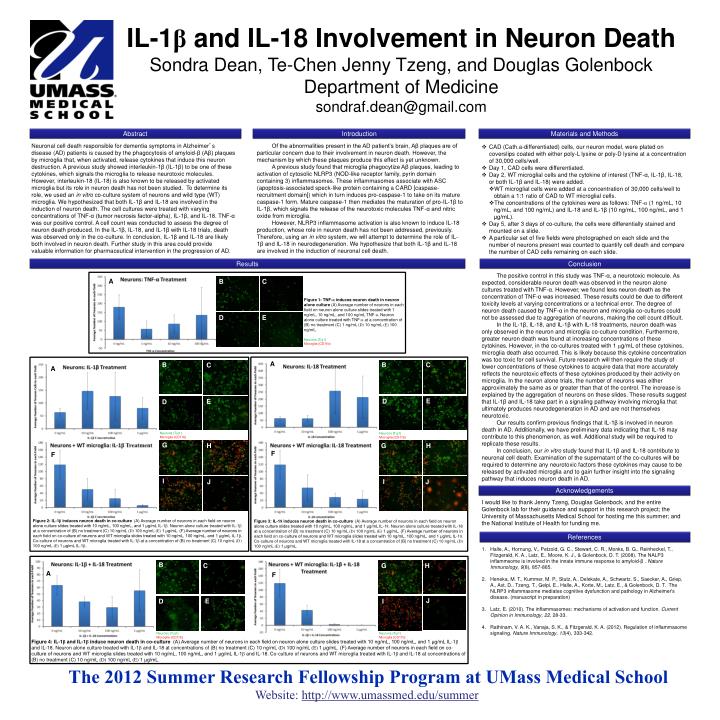
IL-1β and IL-18 Involvement in Neuron Death Sondra Dean, Te-Chen Jenny Tzeng, and Douglas Golenbock Department of Medicine sondraf.dean@gmail.com Abstract Introduction Materials and Methods Neuronal cell death responsible for dementia symptoms in Alzheimer’s disease (AD) patients is caused by the phagocytosis of amyloid-β (Aβ) plaques by microglia that, when activated, release cytokines that induce this neuron destruction. A previous study showed interleukin-1β (IL-1β) to be one of these cytokines, which signals the microglia to release neurotoxic molecules. However, interleukin-18 (IL-18) is also known to be released by activated microglia but its role in neuron death has not been studied. To determine its role, we used an in vitro co-culture system of neurons and wild type (WT) microglia. We hypothesized that both IL-1β and IL-18 are involved in the induction of neuron death. The cell cultures were treated with varying concentrations of TNF-α (tumor necrosis factor-alpha), IL-1β, and IL-18. TNF-α was our positive control. A cell count was conducted to assess the degree of neuron death produced. In the IL-1β, IL-18, and IL-1β with IL-18 trials, death was observed only in the co-culture. In conclusion, IL-1β and IL-18 are likely both involved in neuron death. Further study in this area could provide valuable information for pharmaceutical intervention in the progression of AD. Of the abnormalities present in the AD patient’s brain, Aβ plaques are of particular concern due to their involvement in neuron death. However, the mechanism by which these plaques produce this effect is yet unknown. A previous study found that microglia phagocytize Aβ plaques, leading to activation of cytosolic NLRP3 (NOD-like receptor family, pyrin domain containing 3) inflammasomes. These inflammasomes associate with ASC (apoptosis-associated speck-like protein containing a CARD [caspase-recruitment domain]) which in turn induces pro-caspase-1 to take on its mature caspase-1 form. Mature caspase-1 then mediates the maturation of pro-IL-1β to IL-1β, which signals the release of the neurotoxic molecules TNF-α and nitric oxide from microglia. However, NLRP3 inflammasome activation is also known to induce IL-18 production, whose role in neuron death has not been addressed, previously. Therefore, using an in vitro system, we will attempt to determine the role of IL-1β and IL-18 in neurodegeneration. We hypothesize that both IL-1β and IL-18 are involved in the induction of neuronal cell death. • CAD (Cath.a-differentiated) cells, our neuron model, were plated on coverslips coated with either poly-L lysine or poly-D lysine at a concentration of 30,000 cells/well. • Day 1, CAD cells were differentiated. • Day 2, WT microglial cells and the cytokine of interest (TNF-α, IL-1β, IL-18, or both IL-1β and IL-18) were added. • WT microglial cells were added at a concentration of 30,000 cells/well to obtain a 1:1 ratio of CAD to WT microglial cells. • The concentrations of the cytokines were as follows: TNF-α (1 ng/mL, 10 ng/mL, and 100 ng/mL) and IL-18 and IL-1β (10 ng/mL, 100 ng/mL, and 1 μg/mL). • Day 5, after 3 days of co-culture, the cells were differentially stained and mounted on a slide. • A particular set of five fields were photographed on each slide and the number of neurons present was counted to quantify cell death and compare the number of CAD cells remaining on each slide. Results Conclusion The positive control in this study was TNF-α, a neurotoxic molecule. As expected, considerable neuron death was observed in the neuron alone cultures treated with TNF-α. However, we found less neuron death as the concentration of TNF-α was increased. These results could be due to different toxicity levels at varying concentrations or a technical error. The degree of neuron death caused by TNF-α in the neuron and microglia co-cultures could not be assessed due to aggregation of neurons, making the cell count difficult. In the IL-1β, IL-18, and IL-1β with IL-18 treatments, neuron death was only observed in the neuron and microglia co-culture condition. Furthermore, greater neuron death was found at increasing concentrations of these cytokines. However, in the co-cultures treated with 1 μg/mL of these cytokines, microglia death also occurred. This is likely because this cytokine concentration was too toxic for cell survival. Future research will then require the study of lower concentrations of these cytokines to acquire data that more accurately reflects the neurotoxic effects of these cytokines produced by their activity on microglia. In the neuron alone trials, the number of neurons was either approximately the same as or greater than that of the control. The increase is explained by the aggregation of neurons on these slides. These results suggest that IL-1β and IL-18 take part in a signaling pathway involving microglia that ultimately produces neurodegeneration in AD and are not themselves neurotoxic. Our results confirm previous findings that IL-1β is involved in neuron death in AD. Additionally, we have preliminary data indicating that IL-18 may contribute to this phenomenon, as well. Additional study will be required to replicate these results. In conclusion, our in vitro study found that IL-1β and IL-18 contribute to neuronal cell death. Examination of the supernatant of the co-cultures will be required to determine any neurotoxic factors these cytokines may cause to be released by activated microglia and to gain further insight into the signaling pathway that induces neuron death in AD. A B C Figure 1: TNF-α induces neuron death in neuron alone culture (A) Average number of neurons in each field on neuron alone culture slides treated with 1 ng/mL, 10 ng/mL, and 100 ng/mL TNF-α. Neuron alone culture treated with TNF-α at a concentration of (B) no treatment (C) 1 ng/mL (D) 10 ng/mL (E) 100 ng/mL. D E Neurons (Tuj1) Microglia (CD11b) A B B C C A E D E D Neurons (Tuj1) Microglia (CD11b) Neurons (Tuj1) Microglia (CD11b) G G H H F F I I J J Acknowledgements I would like to thank Jenny Tzeng, Douglas Golenbock, and the entire Golenbock lab for their guidance and support in this research project; the University of Massachusetts Medical School for hosting me this summer; and the National Institute of Health for funding me. Figure 2: IL-1β induces neuron death in co-culture (A) Average number of neurons in each field on neuron alone culture slides treated with 10 ng/mL, 100 ng/mL, and 1 μg/mL IL-1β. Neuron alone culture treated with IL-1β at a concentration of (B) no treatment (C) 10 ng/mL (D) 100 ng/mL (E) 1 μg/mL. (F) Average number of neurons in each field on co-culture of neurons and WT microglia slides treated with 10 ng/mL, 100 ng/mL, and 1 μg/mL IL-1β. Co-culture of neurons and WT microglia treated with IL-1β at a concentration of (B) no treatment (C) 10 ng/mL (D) 100 ng/mL (E) 1 μg/mL IL-1β. Figure 3: IL-18 induces neuron death in co-culture (A) Average number of neurons in each field on neuron alone culture slides treated with 10 ng/mL, 100 ng/mL, and 1 μg/mL IL-18. Neuron alone culture treated with IL-18 at a concentraiton of (B) no treatment (C) 10 ng/mL (D) 100 ng/mL (E) 1 μg/mL. (F) Average number of neurons in each field on co-culture of neurons and WT microglia slides treated with 10 ng/mL, 100 ng/mL, and 1 μg/mL IL-18. Co-culture of neurons and WT microglia treated with IL-18 at a concentration of (B) no treatment (C) 10 ng/mL (D) 100 ng/mL (E) 1 μg/mL. References Halle, A., Hornung, V., Petzold, G. C., Stewart, C. R., Monks, B. G., Reinheckel, T., Fitzgerald, K. A., Latz, E., Moore, K. J., & Golenbock, D. T. (2008). The NALP3 inflammsome is involved in the innate immune response to amyloid-β . Nature Immunology, 9(8), 857-865. Heneka, M. T., Kummer, M. P., Stutz, A., Delekate, A., Schwartz, S., Saecker, A., Griep, A., Axt, D., Tzeng, T., Gelpi, E., Halle, A., Korte, M., Latz, E., & Golenbock, D. T. The NLRP3 inflammasome mediates cognitive dysfunction and pathology in Alzheimer's disease. (manuscript in preparation) Latz, E. (2010). The inflammasomes: mechanisms of activation and function. Current Opinion in Immunology, 22, 28-33. Rathinam, V. A. K., Vanaja, S. K., & Fitzgerald, K. A. (2012). Regulation of inflammasome signaling. Nature Immunology, 13(4), 333-342. B C H G A F J E I D Neurons (Tuj1) Microglia (CD11b) Neurons (Tuj1) Microglia (CD11b) Figure 4: IL-1β and IL-1β induce neuron death in co-culture (A) Average number of neurons in each field on neuron alone culture slides treated with 10 ng/mL, 100 ng/mL, and 1 μg/mL IL-1β and IL-18. Neuron alone culture treated with IL-1β and IL-18 at concentrations of (B) no treatment (C) 10 ng/mL (D) 100 ng/mL (E) 1 μg/mL. (F) Average number of neurons in each field on co-culture of neurons and WT microglia slides treated with 10 ng/mL, 100 ng/mL, and 1 μg/mL IL-1β and IL-18. Co-culture of neurons and WT microglia treated with IL-1βand IL-18 at concentrations of (B) no treatment (C) 10 ng/mL (D) 100 ng/mL (E) 1 μg/mL. The 2012 Summer Research Fellowship Program at UMass Medical School Website: http://www.umassmed.edu/summer