Download

1 / 79
820 likes | 1.11k Views
Chapter 7 BIO 160 Kelly Trainor. Nervous System – Part I. Functions of the Nervous System. Sensory input—gathering information To monitor changes occurring inside and outside the body Changes = stimuli Integration To process and interpret sensory input and decide if action is needed

E N D
Chapter 7 BIO 160 Kelly Trainor Nervous System – Part I
Functions of the Nervous System • Sensory input—gathering information • To monitor changes occurring inside and outside the body • Changes = stimuli • Integration • To process and interpret sensory input and decide if action is needed • Motor output • A response to integrated stimuli • The response activates muscles or glands
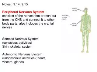
Structural Classification • Central nervous system (CNS) • Brain • Spinal cord • Peripheral nervous system (PNS) • Nerves outside the brain and spinal cord • Spinal nerves • Cranial nerves
Functional Classification of the Peripheral Nervous System • Sensory (afferent) division • Nerve fibers that carry information to the central nervous system • Motor (efferent) division • Nerve fibers that carry impulses away from the central nervous system • Two subdivisions • Somatic nervous system = voluntary • Autonomic nervous system = involuntary
Nervous Tissue: Support Cells • Support cells in the CNS are grouped together as “neuroglia” • Function: to support, insulate, and protect neurons • Astrocytes • Abundant, star-shaped cells • Brace neurons • Form barrier between capillaries and neurons • Control the chemical environment of the brain
Nervous Tissue: Support Cells • Microglia • Spiderlike phagocytes • Dispose of debris
Nervous Tissue: Support Cells • Ependymal cells • Line cavities of the brain and spinal cord • Circulate cerebrospinal fluid
Nervous Tissue: Support Cells • Oligodendrocytes • Wrap around nerve fibers in the central nervous system • Produce myelin sheaths
Nervous Tissue: Support Cells • Satellite cells • Protect neuron cell bodies • Schwann cells • Form myelin sheath in the peripheral nervous system
Nervous Tissue: Neurons • Neurons = nerve cells • Cells specialized to transmit messages • Major regions of neurons • Cell body—nucleus and metabolic center of the cell • Processes—fibers that extend from the cell body • Dendrites—conduct impulses toward the cell body • Axons—conduct impulses away from the cell body
Nervous Tissue: Neurons • Axons end in axonal terminals • Axonal terminals contain vesicles with neurotransmitters • Axonal terminals are separated from the next neuron by a gap • Synaptic cleft—gap between adjacent neurons • Synapse—junction between nerves
Nervous Tissue: Neurons • Myelin sheath—whitish, fatty material covering axons • Schwann cells—produce myelin sheaths in jelly roll–like fashion • Nodes of Ranvier—gaps in myelin sheath along the axon
Neuron Cell Body Location • Most neuron cell bodies are found in the central nervous system • Gray matter—cell bodies and unmyelinated fibers • Nuclei—clusters of cell bodies within the white matter of the central nervous system • Ganglia—collections of cell bodies outside the central nervous system
Functional Classification of Neurons • Sensory (afferent) neurons • Carry impulses from the sensory receptors to the CNS • Cutaneous sense organs • Proprioceptors—detect stretch or tension • Motor (efferent) neurons • Carry impulses from the central nervous system to viscera, muscles, or glands
Functional Classification of Neurons Figure 7.7
Neuron Classification • Interneurons (association neurons) • Found in neural pathways in the central nervous system • Connect sensory and motor neurons
Chapter 7 BIO 160 Kelly Trainor Nervous System – Part II
Functional Properties of Neurons • Irritability • Ability to respond to stimuli • Conductivity • Ability to transmit an impulse
Nerve Impulses • Resting neuron • The plasma membrane at rest is polarized • Fewer positive ions are inside the cell than outside the cell • Depolarization • A stimulus depolarizes the neuron’s membrane • A depolarized membrane allows sodium (Na+) to flow inside the membrane • The exchange of ions initiates an action potential in the neuron
Nerve Impulses Figure 7.9a–b
Nerve Impulses • Action potential • If the action potential (nerve impulse) starts, it is propagated over the entire axon • Impulses travel faster when fibers have a myelin sheath
Nerve Impulses Figure 7.9c–d
Nerve Impulses • Repolarization • Potassium ions rush out of the neuron after sodium ions rush in, which repolarizes the membrane • The sodium-potassium pump, using ATP, restores the original configuration
Nerve Impulses Figure 7.9e–f
Transmission of a Signal at Synapse • Impulses are able to cross the synapse to another nerve • Neurotransmitter is released from a nerve’s axon terminal • The dendrite of the next neuron has receptors that are stimulated by the neurotransmitter • An action potential is started in the dendrite
Transmitting neuron Axonterminal Actionpotentialarrives Axon oftransmittingneuron Vesiclefuses withplasmamembrane Neurotrans-mitter is re-leased intosynaptic cleft Vesicles Neurotransmittermolecules Synaptic cleft Ion channels Synapticcleft Receiving neuron Receivingneuron Synapse Transmission of a Signal at Synapses
Axonterminal Axon oftransmittingneuron Actionpotentialarrives Vesicles Synapticcleft Receivingneuron Synapse Transmitting neuron Neurotrans-mitter bindsto receptoron receivingneuron’smembrane Vesiclefuses withplasmamembrane Neurotrans-mitter is re-leased intosynaptic cleft Neurotransmittermolecules Synaptic cleft Ion channels Receiving neuron Neurotransmitterbroken downand released Neurotransmitter Receptor Na+ Na+ Ion channel opens Ion channel closes Transmission of a Signal at Synapses
Skin Spinal cord (in cross section) Stimulus at distalend of neuron Sensory neuron Integrationcenter Receptor Motor neuron Interneuron Effector (a) The Reflex Arc • Reflex—rapid, predictable, and involuntary response to a stimulus • Occurs over pathways called reflex arcs • Reflex arc—direct route from a sensory neuron, to an interneuron, to an effector
Types of Reflexes and Regulation • Somatic reflexes • Activation of skeletal muscles • Example: When you move your hand away from a hot stove • Autonomic reflexes • Smooth muscle regulation • Heart and blood pressure regulation • Regulation of glands • Digestive system regulation
Chapter 7 BIO 160 Kelly Trainor Nervous System – Part III
Regions of the Brain • Cerebral hemispheres (cerebrum) • Diencephalon • Brain stem • Cerebellum
Regions of the Brain: Cerebrum • Cerebral Hemispheres (Cerebrum) • Paired (left and right) superior parts of the brain • Includes more than half of the brain mass • The surface is made of ridges (gyri) and grooves (sulci)
Regions of the Brain: Cerebrum Figure 7.13a
Regions of the Brain: Cerebrum • Lobes of the cerebrum • Fissures (deep grooves) divide the cerebrum into lobes • Surface lobes of the cerebrum • Frontal lobe • Parietal lobe • Occipital lobe • Temporal lobe
Regions of the Brain: Cerebrum Figure 7.13b
Regions of the Brain: Cerebrum • Specialized areas of the cerebrum • Primary somatic sensory area • Receives impulses from the body’s sensory receptors • Located in parietal lobe • Primary motor area • Sends impulses to skeletal muscles • Located in frontal lobe • Broca’s area • Involved in our ability to speak
Regions of the Brain: Cerebrum Figure 7.13c
Regions of the Brain: Cerebrum Figure 7.14
Regions of the Brain: Cerebrum • Cerebral areas involved in special senses • Gustatory area (taste) • Visual area • Auditory area • Olfactory area • Interpretation areas of the cerebrum • Speech/language region • Language comprehension region • General interpretation area
Regions of the Brain: Cerebrum Figure 7.13c
Regions of the Brain: Cerebrum • Layers of the cerebrum • Gray matter—outer layer in the cerebral cortex composed mostly of neuron cell bodies • White matter—fiber tracts deep to the gray matter • Corpus callosum connects hemispheres • Basal nuclei—islands of gray matter buried within the white matter
Regions of the Brain: Diencephalon • Sits on top of the brain stem • Enclosed by the cerebral hemispheres • Made of three parts • Thalamus • Hypothalamus • Epithalamus
Regions of the Brain: Diencephalon • Thalamus • Surrounds the third ventricle • The relay station for sensory impulses • Transfers impulses to the correct part of the cortex for localization and interpretation
Regions of the Brain: Diencephalon • Hypothalamus • Under the thalamus • Important autonomic nervous system center • Helps regulate body temperature • Controls water balance • Regulates metabolism • An important part of the limbic system (emotions) • The pituitary gland is attached to the hypothalamus • Epithalamus • Forms the roof of the third ventricle • Houses the pineal body (an endocrine gland) • Includes the choroid plexus—forms cerebrospinal fluid
Regions of the Brain: Brain Stem • Attaches to the spinal cord • Parts of the brain stem • Midbrain • Pons • Medulla oblongata
Regions of the Brain: Brain Stem Figure 7.16a
Regions of the Brain: Brain Stem • Midbrain • Mostly composed of tracts of nerve fibers • Has two bulging fiber tracts—cerebral peduncles • Has four rounded protrusions—corpora quadrigemina • Reflex centers for vision and hearing • Pons • The bulging center part of the brain stem • Mostly composed of fiber tracts • Includes nuclei involved in the control of breathing
Regions of the Brain: Brain Stem • Medulla Oblongata • The lowest part of the brain stem • Merges into the spinal cord • Includes important fiber tracts • Contains important control centers • Heart rate control • Blood pressure regulation • Breathing • Swallowing • Vomiting
Regions of the Brain: Brain Stem • Reticular Formation • Diffuse mass of gray matter along the brain stem • Involved in motor control of visceral organs • Reticular activating system (RAS) plays a role in awake/sleep cycles and consciousness