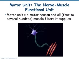
Motor Unit
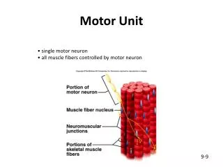
Motor Unit. single motor neuron all muscle fibers controlled by motor neuron. 9-9. Neuromuscular Junction. site where axon and muscle fiber communicate motor neuron motor end plate synaptic cleft synaptic vesicles neurotransmitters. 9-8. Ion Channels.

Motor Unit
E N D
Presentation Transcript
Motor Unit • single motor neuron • all muscle fibers controlled by motor neuron 9-9
Neuromuscular Junction • site where axon and muscle fiber communicate • motor neuron • motor end plate • synaptic cleft • synaptic vesicles • neurotransmitters 9-8
Ion Channels Gated Channels – open and close in response to specific stimuli Chemically (Ligand)Gated Channel – opened and closed by chemicals 2. Voltage Gated Channel – opened and closed based on membrane potential • Mechanically Gated Channel – open and closed by physical deformity • of receptor Leakage Channels – always open
Resting Membrane Potential • occurs when cell is not being stimulated • inside is negative relative to the outside • polarized membrane = electrically charged due to distribution of ions • Na+/K+ pump = pumps Na+ out and K+ in 10-14
Depolarization – reduction in membrane potential -- becomes less negative -- increases probability of impulse conduction Hyperpolarizaiton – increase in membrane potential --becomes more negative -- reduces probability of impulse conduction
Release of Neurotransmitter • Nerve impulse reaches end of the axon and opens Ca2+ channels; Ca2+ • diffuses into the cell • Presence of Ca2+ in axon causes synaptic vesicles to bind with cell • membrane of the axon. • Neurotransmitter is released from axon into synaptic cleft • by exocytosis (Acetylcholine (ACh)) • ACh binds receptors on the motor end plate of the sarcolemma; chemically • gated channels 5. Stimulation of sarcolemma generates an impulse/action potential
Action Potential • Ach opens chemically gated channels on the sarcolemma • Na+ goes into the cell and makes the inside of the membrane positive • relative to the outside • Change in charge inside of the cell opens voltage gated channels • Depolarization moves down the sarcolemma = Action Potential • Membrane repolarizes as ions move back to starting position
Action Potentials 10-18
Excitation Contraction Coupling • muscle impulses travels along the sarcolemma and into T tubule causing terminal cisternae to release Ca2+ • calcium binds to troponin to change its shape • position of tropomyosin is altered • binding sites on actin exposed • actin and myosin bind 9-11
Cross-bridge Cycling • actin and myosin cross-bridge bind • myosin cross-bridge pulls actin • ADP and phosphate released from myosin • new ATP binds to myosin • linkage between actin and myosin cross-bridge break • ATP splits • myosin cross-bridge goes back to original position 9-13 demo
Relaxation • Nerve stops secreting Ach • acetylcholinesterase – breaks down acetylcholine • muscle impulse stops • calcium moves back into sarcoplasmic reticulum • myosin and actin binding prevented 9-14