Download
1 / 15
150 likes | 267 Views
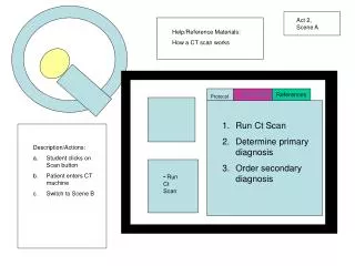
Act 2, Scene A. Help/Reference Materials: How a CT scan works. Protocol. Doctor Notes. References. jjlkjkjlkjkjkj. Run Ct Scan Determine primary diagnosis Order secondary diagnosis. Description/Actions: Student clicks on Scan button Patient enters CT machine Switch to Scene B.

E N D
Act 2, Scene A Help/Reference Materials: How a CT scan works Protocol Doctor Notes References jjlkjkjlkjkjkj • Run Ct Scan • Determine primary diagnosis • Order secondary diagnosis • Description/Actions: • Student clicks on Scan button • Patient enters CT machine • Switch to Scene B • Run Ct Scan
Act 2, Scene B Help/Reference Materials: CT scan of nSAH stroke How to read a CT scan Normal brain CT Protocol Doctor Notes References jjlkjkjlkjkjkj • Run Ct Scan • Determine primary diagnosis • Order secondary diagnosis • Description/Actions: • CT results appear on computer along with diagnostic options • If student chooses SAH stroke, move to Scene C • If student choose nSAH stroke, a window should appear showing what a CT scan of an nSAH stroke looks like • Diagnosis: • A. Subarachnoid Hemorrhagic (SAH) stroke • B. Non-subarchnoid Hemorrhagic (nSAH) stroke
Act 2, Scene C Help/Reference Materials: Definitions/examples of MRI, PET scans, and angiograms Causes of hemorrhagic strokes Protocol Doctor Notes References jjlkjkjlkjkjkj • Run Ct Scan • Determine primary diagnosis • Order secondary diagnosis • Description/Actions: • Student chooses desired test. • If “c”, move to Act 3 • If a or b, a window pops up explaining why this is the wrong answer. • Secondary test options: • MRI • PET scan • CT angiogram
Act 3 • Option 1 • Option 2
Act 3, Scene A, Option 1 Help/Reference Materials: CT angiogram procedure Normal angiograms Protocol Doctor Notes References jjlkjkjlkjkjkj • Perform CT angiogram • Determine course of treatment • Description/Actions: • Student clicks on “run CT angiogram” • CT angiogram is preformed Run CT angiogram
Act 3, Scene B, Option 1 Help/Reference Materials: Aneurysms Clipping vs. coiling Protocol Doctor Notes References jjlkjkjlkjkjkj • Perform CT angiogram • Determine course of treatment Aneurysm in anterior communicating artery • Description/Actions: • CT angiogram results appear • A course of treatment is chosen • Option 2 • Choose a course of treatment: • Clip the aneurysm • Coil the aneurysm
Act 3, Scene A, Option 2 Help/Reference Materials: Catheter information Vascular system anatomy Protocol Doctor Notes References jjlkjkjlkjkjkj • Perform CT angiogram • Determine course of treatment • Description/Actions: • Student clicks on each step and it is done above. • Automatically move to next screen when step 2 is complete. 1. Insert catheter into the femoral artery. 2. Guide catheter through the femoral artery, aorta, and into the vertebral artery.
Act 3, Scene A, Option 2 Help/Reference Materials: Catheter information Vascular system anatomy Protocol Doctor Notes References jjlkjkjlkjkjkj • Perform CT angiogram • Determine course of treatment • Description/Actions: • Student clicks on each step and it is done above. • Automatically move to next screen when step 2 is complete. 1. Inject contrast dye.
Act 3, Scene A, Option 2 Help/Reference Materials: CT angiogram technology Protocol Doctor Notes References jjlkjkjlkjkjkj • Perform CT angiogram • Determine course of treatment • Description/Actions: • Student clicks on “Run CT Angiogram” and machine activates. • Automatically moves to Scene B Run CT Angiogram
Act 3, Scene B, Option 2 Help/Reference Materials: Aneurysms Clipping vs. coiling Protocol Doctor Notes References jjlkjkjlkjkjkj • Perform CT angiogram • Determine course of treatment Aneurysm in anterior communicating artery • Description/Actions: • CT angiogram results appear • A course of treatment is chosen • Choose a course of treatment: • Clip the aneurysm • Coil the aneurysm
Act 3, Scene C Help/Reference Materials: Coiling information Protocol Doctor Notes References jjlkjkjlkjkjkj • Perform embolization (coiling) on the aneurysm. • Send patient to ICU. • Description/Actions: • Student clicks on “coil the aneurysm”. • Coiling procedure is shown. Insert a coil into the aneurysm.
CT scan of nSAH stroke Does your patient’s CT scan match this one? Try again! Your patient’s CT scan Return to ER nSAH Stroke
Correct! The bright white areas indicate a bleed in the subarachnoid space. Great job Dr _______! Proceed! Return to ER SAH stroke Bleeding
Dr. _______’s Notes • Ordered CT Scan • Faster • Can find bleed in brain • Less expensive Return to ER
Dr. _______’s Notes • Ordered CT Scan: faster, can find bleed in brain, less expensive • Subarachnoid hemorrhagic (SAH) stroke diagnosed Return to ER Bleeding