Download
1 / 48
480 likes | 628 Views
Reading Alberts Chapter 8 p. 335-385--Primary reference for nuclear architecture Alberts Chapter 16 p. 800-801--Nuclear Lamins Alberts Chapter 18 p.921-924--Centromeres Alberts Chapter 4 p.139-148--Microscopy Alberts Chapter 4 p. 186-188--Antibody labeling. Alberts 4-20. Nucleus

E N D
Reading • Alberts Chapter 8 p. 335-385--Primary reference for nuclear architecture • Alberts Chapter 16 p. 800-801--Nuclear Lamins • Alberts Chapter 18 p.921-924--Centromeres • Alberts Chapter 4 p.139-148--Microscopy • Alberts Chapter 4 p. 186-188--Antibody labeling
Nucleus • Storage of genetic information • Replication of genetic information • Transcription of genetic information into “functional forms” • Control of gene expression Alberts 1-18
Prokaryotic cells • No nucleus • DNA localizes to nucleiod body • Lack of compartments Alberts 1-12
Replication, Transcription and Translation occur in the cytoplasm of prokaryotic cells • Mechanisms of control are different than in eukaryotic cells. Alberts 3-15
Active transcription and translation in E. coli • Coupled events • Ribosomes attach to transcript while it is still being synthesized Voet 29-18
Eukaryotic cells • Compartmentalized • Replication, transcription and processing occur in the nucleus • Translation occurs in the cytoplasm • Mitochondrial protein synthesis Alberts 3-15
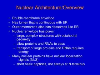
Nuclear organization • Nuclear envelope • Nuclear lamina • Nuclear pore • Nucleolus • Heterochromatin • Euchromatin Alberts 8-1
Electron micrograph of nucleus Alberts 8-71
Nucleus • Nuclear envelope • Lipid bilayer • Permeable to small nonpolar substances • Inner membrane • Continuous with outer membrane • Associates with nuclear lamins • Intermembrane space • Closely resembles ER • Outer membrane • Continuous with lumen of ER Cytoplasm Voet 11-13
Nuclear pores • Major connection between nucleus and cytoplasm • Active and passive transport • Nuclear Lamina • Meshwork of intermediate filament protein lamin • Major structural support for nucleus Alberts 16-18
Nuclear Lamina • Grid-like structure • Composed of lamins A, B, C • Assembly dependent on phosphorylation • Lamin B attaches to inner membrane Alberts 16-18
Nucleolus • Euchromatin • Heterochromatin • Constitutive • Facultative Cooper 8-15
Metabolic labeling with tritiated uridine to identify areas of active transcription • Euchromatin • Heterochromatin Alberts 8-26
Spliceosomes May function as storage centers for splicing factors Or may be sites of splicing Contain splicing factors Speckles PML bodies Disrupted in acute premyelocytic leukemia Associated with nuclear matrix Function unknown Coiled bodies Cajal bodies or Nucleolar accessory bodies Ramon y Cajal described in 1903 May be involved in snRNP production Gems Associated with coiled bodies (Gemini of coiled body Similar in structure to CB, but contains different proteins Other Nuclear Structures
Antibodies are important tools for identification and localization of proteins in cells • Primary antibody • Secondary antibody • Fluorescent, gold or enzymatic label detects antibody labeling indirectly Alberts 4-64
Fluorescence Microscopy • Path • Confocal • Two photon • Deconvolution • Excitation • Emission Alberts 4-7
What is a fluorophore? • Heterocyclic compounds • Absorption of light raises energy to an excited state • Molecule decays to its ground state by emitting photons Alberts 4-8
Other fluorochromes • Dapi and Hoechst stain nucleic acids • Mito-trackers designed to stain specific organelles (mitochondria, lysosomes, etc). • GFP--jellyfish green fluorescent protein • cDNA isolated in 80’s by Bill Ward • Protein folds into a fluorochrome • Make chimeric genes with GFP cDNA and your cDNA • Fluorescent tag that can be visualized in live cells
Fluorescent staining of a fibroblast nucleus • Blue: DNA stained with Dapi • Red: RNA stained with rhodamine labeled poly dT • Green: Fluorescein labeled antibody to a protein involved in splicing Lodish 11-21
GFP fusion proteins expressed in the nucleus • Phair and Misteli (2000) Nature 404: 604-609 Figure 1
FRAP Fluorescence recovery after photobleaching High intensity pulse of laser Fluorochrome hit by laser is dead Monitor the fluorescence over time Recovery of fluorescence due to movement of new molecules into the area Measures movement FRET Fluorescence resonance energy transfer Requires 2 fluorochromes Emission of one must overlap excitation of the second Close proximity of labeled components allows energy transfer Measures proximity of components
We have 2 m of DNA in every nucleus in our body. • Each nucleus is only 10 µm in diameter. • How does all this DNA get packed into such a small space? • Answer: dense packing of the DNA into chromatin. Alberts 18-14
A. Native structure of condensed chromatin • 30 nm fiber • B. Structure of “decondensed” chromatin • Beads on a string • Kornberg 1974 Alberts 8-9
Kornberg’s experiment • Limited digestion of DNA with nuclease release DNA fragments 200 bp long (string). • Further digestion released nucleosome beads. • Dissociation of beads revealed 148 bp of DNA and histones Alberts 8-10
Histones • Small, highly basic proteins (20-30% lys or arg) • Highly conserved • Core histones • Nucleosomal histones • H2A, H2B, H3, H4 • Two copies per nucleosome. • H1 histones • 6 highly related species Alberts 8-10
Model for the structure of the nucleosome. • DNA is sharply bent. • Spool like structure 11 nm in diameter. • Core histones possess similar folds. Alberts 8-10
H1 Histones • Associates with the nucleosome and additional DNA • Chromatosome • Interacts with H1 histone of adjacent chromatosome Alberts 8-15
Interaction of H1 histones with adjacent H1 histones produces the 30 nm structure known as the solenoid. Cooper 4-10
Radial Loops • EM of isolated chromosomes display loops of chromatin extending from a backbone • Lampbrush chromosomes also display loops • More extensive folding • Scaffold for chromosome structure Alberts 8-29
Double helix • Nucleosomes • Solenoids • Radial loops • Condensed loops • Metaphase chromosome • Heterochromatin may closely resemble metaphase chromosome • Euchromatin may be structurally similar to 10 nm/30 nm structures Alberts 8-30
Scaffolds • Extensive radial loops appear to extend from a backbone • Removal of histones allows observation of what appears to be a fibrous core or scaffold • Real or Artifact? • Harsh treatment may cause deposition of protein in the sticky nucleic acid Voet 33-14
How does chromatin structure effect replication and transcription?
Model for dealing with histones during replication • Nucleosome splits in half as the replication fork approaches • Histones remain associated with one strand • Nucleosome re-forms after replication passes • New histones are added to other pair Alberts 8-40
Cooper 6-32 • Activation • Acetylation of histones correlates with actively transcribed genes • Acetylation reduces the net positive charge of the histones • HMG14 or 17 compete with H1 histones for binding to the nucleosome • De-acetylation is involved in turning transcription off
Metaphase chromosome • Chromatids • Telomeres • Centromere • Holds sister chromatids together • Attachment of kinetochore during mitosis Alberts 18-15
Origins of replication-sites where DNA replication begins. • Telomeres are specialized sequences at the end of linear chromosomes that ensure that genetic material is not lost during replication. • Centromeres hold chromatids together prior to cell division and associate with kinetochores. Alberts 8-4
Kinetochore • Attachment site for spindle microtubules • DNA/ protein structure • Required for proper chromosome segregation Alberts 18-16
Yeast centromere has been characterized. • Simple DNA sequence that binds to microtuble Alberts 18-17
Chromosome Banding • Chromomeres • Distinctive banding pattern for each chromosome • Hoechst (G bands--AT rich sequence) • Olivomycin (R bands--GC rich sequence) • Giemsa • Feulgen reagent Alberts 8-31
Domains of replication • Metabolically label cells with bromodeoxyuridine • Substitutes for thymidine • Alters staining of G bands or can use antibodies to detect • Synthesis occurs in distinct domains during S phase Alberts 8-37
In situ hybridization • Examine position of a gene on a chromosome • Examine position of chromosome in nucleus • Examine distribution of transcript in cytoplasm • Basic hybridization technique • Label probe • Hybridize via base pairing to target Alberts 7-17
Result of in situ hybridization • Gene specific probes • Unique labels for each gene • Metaphase chromosome 5 • Duplicate spots for each probe • Positions on each pair similar Alberts 7-19