
Case of Fulminant Invasive Pneumococcal Disease in a Middle-Aged Patient with Sickle Cell Trait
This case study examines a 49-year-old male with no previous comorbidities who presented with severe invasive pneumococcal disease (IPD), exhibiting symptoms such as fever, multi-organ failure, and confusion. Notably, he was found to have sickle cell trait, which is a rare but significant association with increased risk of acute infections and complications. His case underscores the importance of considering underlying hematological conditions in patients experiencing invasive pneumococcal infections. The patient required intensive care and was discharged with ongoing prophylaxis against pneumococcal infections.

Case of Fulminant Invasive Pneumococcal Disease in a Middle-Aged Patient with Sickle Cell Trait
E N D
Presentation Transcript
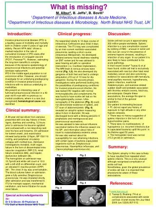
What is missing?M. Albur2, B. Jeffs1, B. Bovill11Department of Infectious diseases & Acute Medicine, 2Department of Infectious diseases & Microbiology, North Bristol NHS Trust, UK Introduction: Clinical progress: Discussion: Invasive pneumococcal disease (IPD) is a leading cause of morbidity and mortality both in children under 5 years of age and elderly. Recent HPA data1 shows a marked reduction in the rate of cumulative increase of IPD cases after the introduction of conjugate vaccine (PCV7, PrevenarTM). However, estimating the long-term benefits is complex because of the dynamic epidemiology of pneumococcal infections and potential serotype replacement. IPD in the middle-aged population is not uncommon either. However, one should investigate for an underlying predisposing condition in invasive pneumococcal infections with no obvious underlying co-morbidities. We present an interesting case of fulminant pneumococcal infection in a 49 year old fit man with no previous co-morbid conditions, where in a rare (but recognized) hematological cause was identified • Sickle cell trait occurs in approximately 300 million people worldwide. Splenic infarction is a rare complication caused by sickling of RBC - unusual in sickle cell trait, but can occur in the presence of extreme hypoxic stresses, resulting in infarction of any organ. In our patient it is also likely to have contributed to his acute pathology. • In their recent review2 Tsaras G et al have found, sickle cell trait is exclusively associated with rare but often fatal renal medullary cancer and also convincing evidence for associations with hematuria, renal papillary necrosis, hyposthenuria, splenic infarction, exertional rhabdomyolysis, and exercise-related sudden death and probable association with thrombo-embolic events, fetal loss, pre-eclampsia. Despite these associations, the average life span is similar to that of the general population. • Our patient is interesting because • Pt. did not report any history of exposure to high altitude or any other hypoxic stresses. • There was no history suggestive of splenic infarction in the form of left hypochondriac pains. • There were no manifestations of hyposplenia (i.e. recurrent infections due to capsulated bacteria) uptill this point in his lifetime aged 50 years. • The hemoglobin level was normal • (15.2 gm/dl) on admission. He responded slowly to 14 days course of intravenous Ceftriaxone plus initial 4 days of steroids. The ITU stay was complicated by an inter-current ventilator-associated pneumonia needing a short course intravenous Meropenam. He developed bilateral sensory-neural deafness needing an ENT review and he was advised to wear hearing aid with no operative intervention (i.e. Cochlear implantation). The MRI brain & venous sinuses were normal (Fig. 1,2). He also developed dry gangrene of both feet and had to undergo amputation of 8 out of 10 toes for dry gangrene. During his recovery phase, investigations were performed looking into the possibilities of predisposing conditions for invasive pneumococcal infection. He was tested HIV negative with normal immunoglobulins and complements. He was found to have sickle cell trait on his hemoglobinopathy screen. Ultra-sonography of the abdomen (Fig. 3) could not demonstrate existence of spleen, and CT scan of abdomen/pelvis (Fig. 4) revealed an atrophic spleen measuring about 4.6cm in diameter. He was discharged home with a lifelong penicillin V prophylaxis and meningococcal and pnemococcal vaccinations. He was advised to take annual influenza vaccination before the influenza season via his GP, and information about risks of travel to malaria/babesia endemic areas. He was also warned about his susceptibility for over whelming infections with certain types of bacteria (capsulated organisms such as Streptococcus pneumoniae, Haemiphilus influenzae, and Capnocytophaga canimorsus etc) Clinical summary: A49-year-old taxi-driver from Jamaica presented with one-day history of fever, rigors, diarrhea and vomiting. 12 hours prior to admission he became agitated and confused with a non-blanching rash over his face and forearms. On admission he looked unwell, and examination showed tachycardia 121bpm, BP 150/110 mmHg, and peripheral circulatory collapse with mottled toes. Admission investigations revealed, multi-organ failure in the form of disseminated intra-vascular coagulation (INR 2.5), renal failure (creatinine 489), and hepatic dysfunction (billirubin 98). The hemoglobin on admission was 15.7gm/dl and white cell count of 12.0 with a left shift on differential count, and an elevated C-reactive protein (CRP) 281. The chest X-ray was unremarkable . The blood cultures taken on admission grew a fully sensitive Streptococcus pneumoniae. He was intubated shortly after admission and was transferred to ITU for inotropic support, mechanical ventilation, and hemo-filtration for acute renal failure. Summary: The Splenic atrophy in this case is likely to have resulted from repeated silent splenic infarcts. This is a very unusual although recognised complication of sickle cell trait. Nonetheless, given the global prevalence of sickle cell trait, it is important that physicians be aware of these associations. Fig. 1 Fig. 2 References: Fig. 4 Fig. 3 1.www.hpa.org.uk 2. Tsaras G, Boateng FA et al complications associated with sickle cell trait- A brief review Am Jour Med 2009 Jun;122(6):507-512. Special acknowledgements to: Dr S G Glover, ID Physician & ITU Staff at North Bristol NHS Trust


