1 / 23
230 likes | 291 Views
Erwin Chargaff was one of a handful of scientists who expanded on Levene's work by uncovering additional details of the structure of DNA, thus further paving the way for Watson and Crick. Chargaff, an Austrian biochemist, had read the famous 1944 paper by Oswald Avery and his colleagues at Rockefeller University, which demonstrated that hereditary units, or genes, are composed of DNA. This paper had a profound impact on Chargaff

E N D
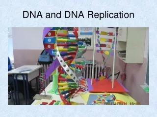
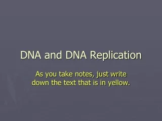
Watson and Crick model of DNA Some Basic • Nucleoside- is a compound formed by the combination of a pentose sugar and nitrogen base. • Nucleotide-is a compound formed by the combination of nucleoside and phosphate group. • Nucleotides building blocks of nucleic acids. • Nucleotide have three characteristic components. • A nitrogenous base • A pentose sugar • A phosphate group
Nitrogen bases present in DNA • Adenine 3.Cytosine • Guanine 4.Thyrosine The different types of nucleosides of DNA • Deoxy adenosine 3.Deoxy cytidine • Deoxy guanosine 4.Deoxy tymidine The DNA contain 2 major purine bases • Adenine • Guanine The DNA contain 2 pyrimidine bases • Cytosine • Thymine
X ray diffraction • x-ray crystallography was originaly used to look at the structures of simple organic minerals,but also progressively applied to more and complex molecules. • It aided in determining the structures of the alpha helix,the beta sheets, hemoglobin and DNA.
DNA is double helix • To shed more light on the structure of DNA. Rosalind, Franklin and wilkins used the powerful method of X-ray diffraction to analyses DNA fibres. • They showed in the early 1950s that DNA produces a charectestic X-ray diffraction pattern • From this pattern it was deduced that DNA molecules are helical with two periodicities along their along axis • A primary one is 3.4A and a secondary one is 34A • The problem then was to formulate a three dimensional model of DNA molecules that could account not only for X-ray diffraction data but also for the specific A=T and G=C base.equivalence discovered by chargaff and for the other chemical properties of DNA
X-ray diffraction and the structure of DNA Watson was shown this picture by wilkins in early 1953 from this picture it was possible to calculate • The distance between base (3.4A) • The length of the period (34A) • The rise of the helix (36degree)
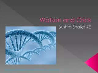
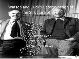
Watson and Crick model of DNA • In 1953 watson & crick proposed a three dimensional model of DNA Structure • It consists of two helical DNA chains wound around the same axis to form a right handed double helix. • The hydrophilic back bones of alternating deoxy- ribose and phosphate groups are on the outside of the double helix . • The purine and pyrimidine bases of both strands are stacked inside the double helix. • The hydrophobic planar ring structure very close together and perpendicular to the long axis
The offset pairing of two strands creates a major groove and minor groove on the surface of the duplex. • Each neucleotide base of one strand is paired in the same plane with a base of the other strand. • According to this model DNA is antiparallel it is ultimately confirmed by x-ray analysis. • The distance between successive base pairs is 3.4A. • The every turn of the helix measures 34A and contain 10 base pairs.
The steps are formed by paired nitrogen base • A always pairs with T with 2 hydrogen bonds and G pairs with C with 3 hydrogen bonds • Hence two strands are complementary to each other • Because of the specificity in base pairing the amount of purines is equal to the amount of pyrimidines A=T & G=C • This is called chargaff`s rule of base equivalence. • The DNA double helix is held together by two forces,hydrogen bonding between complementary base pairs and base stacking interactions. • These two force gives the stability of the double helix.
For their outstanding work in discovering the double helical structure of DNA • Watson and crick shared the 1962 Nobel price for physiology and medicine with wilkins,sadly and franklin whose working greatly contributed to this key discovery.
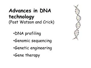
The discovery of the structure of DNA by watson and crick in 1953 • It was a momentous event in science • An event gave rise to entirely new disciplines and influenced the course of many established ones • Our precise understanding of the storage and utilization of cells genetic information is based on work made possible by this discovery
If you would like to donate us? Scan below and donate us 0.013$ (US dollar) (5Rs Indian rupee) Contact: If you want PPT/PDF files, please contact below. Email: gnccmysore@gmail.com Telegram:+919738137533(only for Chat)