STEVEN J. ZEHREN, PH.D.
350 likes | 974 Views
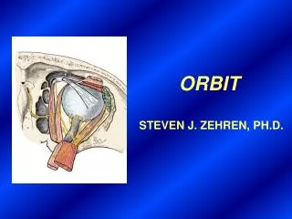
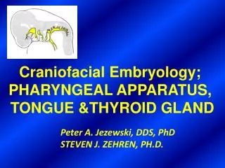
EMBRYOLOGY OF THE PHARYNGEAL APPARATUS, TONGUE &THYROID GLAND. STEVEN J. ZEHREN, PH.D. PHARYNGEAL APPARATUS. Site of midbrain. Pharyngeal (branchial) arches. Lens placode. Somites. Max. prom. 4. 3. 2. 1. Nasal placode. Mand. prom. Stomodeum. Heart. A.

STEVEN J. ZEHREN, PH.D.
E N D
Presentation Transcript
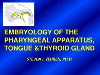
EMBRYOLOGY OF THE PHARYNGEAL APPARATUS, TONGUE &THYROID GLAND STEVEN J. ZEHREN, PH.D.
PHARYNGEAL APPARATUS
Site of midbrain Pharyngeal(branchial) arches Lens placode Somites Max. prom. 4 3 2 1 Nasal placode Mand. prom. Stomodeum Heart A PHARYNGEAL ARCHES FORM DURING 4TH WEEK. ECTODERMAL GROOVES SEPARATE ARCHES ON OUTSIDE. Slide 10.1
STOMODEUM & OROPHARYNGEAL MEMBRANE Endoderm of pharynx Oropharyngeal membrane Ectoderm Stomodeum (primitive oral cavity)
Frontonasal prominence Eye Maxillary prominence Entrance to stomodeum Mandibular prominence Second pharyngeal arch Umbilical vein Third pharyngeal arch Fourth pharyngeal arch Entrance to intraembryonic coelom Spinal cord SCANNING EM OF EMBRYO (5TH WEEK) Slide 10.31
Mesoderm Endoderm Ectoderm 1st pharyngeal pouch Aortic arches (1st to 4th) Midbrain Esophagus Level ofsection C Lung bud Dorsal aorta Esophagus B Thyroid diverticulum (primordium of thyroid gland) Heart Truncus arteriosus (common arterial trunk from heart) Germ Layer Derivatives ENDODERMAL PHARYNGEAL POUCHES SEPARATE ARCHES ON INSIDE Slide 10.2
Mesoderm Endoderm Ectoderm Mandibular (1st) arch 1st arch (Meckel) 1st pharyngeal (branchial) membrane Cartilages 2nd arch (Reichert) Hyoid (2nd) arch Nerve 2nd pharyngeal pouch Muscle 3rd aortic arch C 3rd pharyngeal pouch Mesodermal core of 4th arch Germ Layer Derivatives COMPONENTS OF A PHARYNGEAL ARCH Slide 10.3
ZYGOMATIC MAXILLA MANDIBLE SQUAMOUS TEMPORAL MEMBRANE BONES OF ARCH 1
(Arch 2) (Arches 4 &6) (Arch 1) (Arch 1) (Arch 3) PHARYNGEAL ARCH NERVES
3rd, 4th, and 6th aortic arches Spinal cord Midbrain Pulmonary artery Dorsal aorta Aortic sac Yolk stalk Yolk sac C PHARYNGEAL ARCH ARTERIES (AORTIC ARCHES) Slide 14.56
6th aortic arch 3rd aortic arch 4th aortic arch Truncus arteriosus Dorsal aortae Aortic sac Left dorsal aorta External carotid artery Left dorsal aorta Internal carotid artery Right 3 3 4 Aortic sac 4 5 Aortic arches 5 6 6 Ductus arteriosus Truncus arteriosus (partly divided aortic and pulmonary arteries) Right subclavian artery Left dorsal aorta Aortic sac Pulmonary arteries Left subclavian artery 7th intersegmental artery 7 WKS 6 WKS A B TRANSFORMATION OF AORTIC ARCHES (VENTRAL VIEW) Slide 14.57
3rd aortic arch 6th aortic arch 4th aortic arch Aortic sac Dorsal aortae Truncus arteriosus Internal carotid arteries Left common carotid artery External carotid arteries Right Brachiocephalic artery Left subclavian artery Subclavian arteries Arch of aorta Ascending aorta Right pulmonary artery Ligamentum arteriosum Ductus arteriosus Left pulmonary artery Ascending aorta Descending aorta C D Pulmonary trunk 8 WKS 6 MO. INFANT TRANSFORMATION OF AORTIC ARCHES (VENTRAL VIEW) Slide 14.58
PHARYNGEAL POUCHES, GROOVES & MEMBRANES Opercular flap A B
Auditory tube(pharyngotympanic tube) and tympanic cavity (pouch I) Foramen cecum of tongue Tonsillar sinus and surface epitheliumof palatine tonsil(pouch II) Tongue Tract ofthyroglossal duct Larynx Pouch IV Parathyroidglands Pouch III Ultimobranchialbody (pouch IV) Thyroid gland Thymus (pouch III) 20 WEEK FETUS SHOWING DERIVATIVES OF PHARYNGEAL POUCHES Slide 10.13
Hyoid bone Undescended parathyroid gland Accessory thymic tissue Persistent thyroglossal duct Thyroid cartilage Thyroid gland Superior parathyroid glands Trachea Persistent cord of thymic tissue Manubrium of sternum Ectopic inferior parathyroid gland Retrosternal thymus Body of sternum CONGENITAL ANOMALIES OF THYROID, THYMUS & PARATHYROID GLANDS Slide 10.19
LATERAL CERVICAL CYSTS (REMNANTS OF CERVICAL SINUS) Epicardial ridge (SCM develops in it) C A B
LATERAL CERVICAL CYST. NOTE ITS POSITION ANTERIOR TO SCM MUSCLE. Sternocleidomastoidmuscle Swelling formed bybranchial cyst Tendon ofsternocleidomastoidmuscle Slide 10.16
Distal tongue bud Arches: Migration of thirdarch mesoderm 1 Median tongue bud 2 Hypobranchialeminence Foramen cecumof tongue 3 4 Copula Rima glottidis(opening to vocalapparatus) Hypobranchialeminence A B 5 WKS Esophagus Laryngotracheal groove 4 WKS Median sulcus Oral part of tongue Circumvallatepapillae Terminal sulcus Pharyngeal part of tongue Foramen cecum of tongue C Epiglottis ADULT Arch Derivatives of Tongue 2nd pharyngeal arch (CN VII-chorda tympani) 1st pharyngeal arch (CN V-mandibular division) 3rd pharyngeal arch(CN IX-glossopharyngeal) 4th pharyngeal arch(CN X-vagus) DEVELOPMENT OF TONGUE Slide 10.26
Temporalis Orbicularis oculi Frontalis Auricularis Occipital myotomes Buccinator Occipitalis Stylohyoid Orbicularis oris Stylopharyngeus Masseter Anterior and posterior bellies of digastric muscle Mylohyoid Pharyngealmuscles Platysma B A Clavicle Sternocleidomastoid First archmuscles Second archmuscles Third archmuscles Fourth and sixtharch muscles ALL TONGUE MUSCLES (EXCEPT PALATOGLOSSUS) DEVELOP FROM OCCIPITAL MYOTOMES Slide 10.9
Pharyngeal pouches Primordialpharynx Thyroiddiverticulum 4 WKS A Esophagus Pharyngealarches Heart Laryngotrachealdiverticulum Oropharyngealmembrane Foramen cecum of tongue Tongue Thyroglossal duct Thyroid diverticulum 5 WKS Esophagus B Stomodeum Former site oforopharyngealmembrane Developinghyoid bone DEVELOPMENT OF THYROID GLAND (EARLY) Slide 10.20
Foramen cecum Thyroglossal duct Site of atrophyof duct C Trachea 6 WKS Thyroid gland Hyoid bone Tongue Soft palate Hard palate Foramen cecumof tongue Former tract ofthyroglossal duct Hyoid bone Larynx Pyramidal lobeof thyroid gland Thyroid gland ADULT D DEVELOPMENT OF THYROID GLAND (LATER) Slide 10.21
Foramen cecum of tongue Lingualthyroglossalduct cyst Hyoid bone Hyoid bone Thyroid cartilage Thyroglossal duct cyst Thyroid gland A B Cervical thyroglossal duct cyst Opening of thyroglossal duct sinus THYROGLOSSAL DUCT CYSTS AND FISTULA (SINUS) NOTE MIDLINE POSITION OF CYSTS Slide 10.22