Download
1 / 52
520 likes | 687 Views
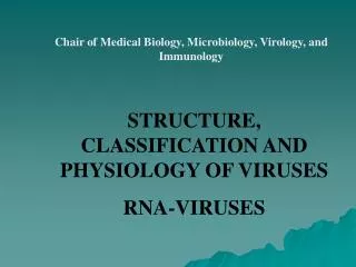
C hair of Medical Biology, M icrobiology, V irology, and I mmunology. Rubella virus Rhabies virus Rotaviruses Coronaviruses. Lecturer As. Prof. O.Pokryshko. Rubellaviruses. The rubella virus is a member of the genus Rubivirus in the family Togaviridae. Rubella.

E N D
Chair of Medical Biology, Microbiology, Virology, and Immunology Rubella virus Rhabies virus Rotaviruses Coronaviruses Lecturer As. Prof. O.Pokryshko
Rubellaviruses The rubella virus is a member of the genus Rubivirus in the family Togaviridae.
Rubella (German measles) is a common mild disease characterized by a rash. It affects children and adolescents worldwide and can also affect young adults. When rubella virus infects susceptible women early in pregnancy, it may be transmitted to the fetus and may cause birth defects. Therefore, accurate diagnosis is critical in pregnancy.
Cataract a child born with cataracts as a result of CRS (Congenital Rubella Syndrome)
Shape: bullet • Genome: -ssRNA • enveloped virus • CPE: Negri body
Reservoir • Urban forms: Dogs and cats • Sylvatic forms: Bats, foxes, raccoons, wolves, skunks, coyotes, mongooses, and biting animals
Transmission • By animal bite or scratch • Via saliva • Airborne ( bats)
Transmission of Rabies • Usually thru infected saliva entering bite wounds. Virus migrates up peripheral nerves to the spinal cord and ends up in the brain. • Aerosol transmission has been documented from caves with large populations of infected bats. • Organ donations—documented from early corneal transplantation. In 2004 four US citizens died from rabies acquired via organ transplantation from the same donor.
Incubation of Rabies • Averages three to eight weeks • Can be as short as 1 week or up to 1 year • Bite location and amount of virus present are the two most important factors in incubation of the virus. • Virus migrates from peripheral nerves to the spinal cord and ends up in the brain. The Virus replicates in neurons and migrates out of the brain into the salivary glands
Clinical Signs of Rabies • Normally has 3 defined stages • 1. Prodromal Phase—Behavioral changes This stage lasts 1-3 days • A. Friendly animals become shy and fractious • B. Wild animals loose their fear of humans • C. Nocturnal animals come out in the day
Clinical Signs of Rabies • 2. Hyperactive Stage It lasts 1-4 days. • A. Easily Excited • B. Bite anything close by • C. Bite imaginary objects • In some cases the animal does not exhibit the Hyperactive stage. These animals appear to be in a stupor. This is called “dumb” rabies
Clinical Signs of Rabies 3. Paralytic Stage • Viral damage to motor neurons results in paralysis. This is usually seen first in the hind legs. In coordination is one of the first signs of the paralytic stage of Rabies. • Paralysis of the throat causes drooling and the inability to swallow. Loss of Jaw tone, dropped Jaw. This stage lasts 1-2 days and is followed by death due to respiratory failure
Rabies Virus • Virus multiplies in skeletal muscles and then brain cells, causing encephalitis • Initial symptoms may include muscle spasms of the mouth and pharynx and hydrophobia
Prevention of Rabies • Preexposure prophylaxis: Injection of human diploid cells vaccine (HDCV) • Postexposure treatment: Vaccine plus rabies immune globulin (RIG)
No effective treatment exists. Postexposure Prophylaxis/PEP: 3 steps 1. Wound care: immediate thorough washing with soap and water and a virucidal agent such as povidine-iodine or 1-2% benzalkonium chloride. • Shown to be protective if performed within 3 hours of exposure • If puncture, swab deeply in wound and around edges
2. Passive Immunization: Human rabies immunoglobulin (HRIG) 20 IU/kg ASAP, but not longer than 7 days after vaccine given. Infiltrate entire dose around wound, any remaining IG inject IM at a site distant from the vaccine. • 3. Human diploid cell vaccine (HDCV): 1 ml (deltoid) on days 0,3,7,14,28. HRIG and HDCV: give in different anatomical sites and never in the same syringe
Challenge Virus Standard • Fatal encephalo-myelitis • Acute rabies • Pasteur Virus strain • Abortive rabies (myelitis) with paralytic sequelea • Abortive rabies
Rabies vaccine types • Human Diploid Cell Vaccine (HDCV) • Rabies Vaccine adsorbed (RVA) • Purified chicken embryo cell (PCEC) Red Book 2003
Laboratory diagnosis • Diseased dog: viral antigen and Negri body in brain tissue. • Patient: IF assay, PCR.
Diagnosing Rabies • 1.Negri Bodies • Italian Physician discovered them in 1903. • Cytoplasmic inclusion bodies in neurons • Rapid and easy to do in the lab Neuron with Negri body
Diagnosing Rabies • 2. Mouse Inoculation Test • Very accurate • Disadvantage is it can take 2 to 3 weeks to make the final conclusion. • Not in common use due to the accuracy of the florescent antibody test
Diagnosing Rabies • 3. Fluorescent Antibody Test • 99%+ accurate • Postmortem on brain tissue • Different Serotypes are present, approximately 8 virus variants • PRIMARY METHOD • USED TODAY
Immunofluorescent viral inclusions in a peripheral nerve in a cryostat section from a patient with rabies, obtained via an antemortem nuchal skin biopsy. Rupprecht CE, The Lancet Infectious Diseases Vol 2 June 2002
Rotavirus structure • Naked double shell capsid • 11 segment double stranded RNAgenome • Replication in cytoplasm
Rotaviruses infections • Account for 50-80% of all cases of viral gastroenteritis. • Usually endemic, but responsible for occasional outbreaks. • Causes disease in all age groups but most severe symptoms in neonates and young children. • Asymptomatic infections common in adults and older children. Symptomatic infections again common in people over 60. • Up to 30% mortality rate in malnourished children, responsible for up to half a million deaths per year. • More frequent during the winter.
Rotavirus / Pathogenesis • Fecal oral transmission, fomites • Highly infectious, 90% of children are seropositive by age 3 • 1012 particles/ml in stool; infection can result from 10 particles • Incubation period 1-3 days • 24-48 hr incubation period followed by an abrupt onset of vomiting and diarrhoea, a low grade fever may be present.
Rotavirus Clinical Features • Fever, vomiting, watery diarrhea dehydration • No blood or leukocytes in stool • Virus replicates in epitheal cells of villi in small intestine • Damage to epithelium major cause of diarrhea • One virus gene product is an enterotoxin • Causes loss of electrolytes and prevents readsorption of water • Self limiting; can be fatal in malnourished or dehydrated children Gastrointestinal symptoms generally resolve in 3 to 7 days
Rotavirus Complications • Severe diarrhea • Dehydration • Electrolyte imbalance • Metabolic acidosis • Immunodeficient children may have more severe or persistent disease
Clinical appearance of dehydrationPhoto Credit: Dr. D. Mahalanabis, World Health Organization
DIAGNOSIS Antigen detection in stool ELISA, LA (Group A rotavirus), immunochromatographic assay Culture- Group A rotaviruses can be cultured in monkey kidney cells Serologyfor epidemiologic studies
Treatment and prevention Treatment Supportive- oral, IV rehydration Prevention Hand hygiene and disinfection of surfaces Vaccine Live oral vaccine
Coronavirus • Viral genome structure: • The non-segmented RNA genome • The virions is about 100 to 140 nm in diameter • Viral protein structure: • Pleomorophic and enveloped particles • Characteristic long projected surface protein (spike) about 20 nm long www.cell-research.com/ 20033/sars.jpg
Transmission • SARS-CoV is predominantly spread in droplets shed from respiratory secretions of patients direct or indirect contacts • Less likely by oral-fecal transmission fecal or airborne transmission • Viral load is peaked at around 10 days and 13 -14 days in stool
Symptoms • Clinical course: • short incubation period (6 days) • time period from exposure to onset of symptoms ranging from 2 to 16 days • intensive care usually required about 10 days after onset of symptoms • There are generally 3 phases: • week 1: cold like symptoms,fever, myalgia, chill and a sore throat • week 2, recurrence of fever. Onset of diarrhea, and oxygen desaturation • only 20% of patients reach this phase, requires ventilatory support • The fatality rate is about 10% upon infection
Chills and shivering & muscle aches Breathing difficulties 3-7 days from onset of symptoms Sudden onset of high fever (>38°C) & dry cough www.health24.com/ images/center/sars_flu.jpg Symptoms
Antiviral drugs/Vaccines • Currently, no specific antiviral drugs available for SARS-CoV • Vaccines are under development: • In China, a second-phase human trials of a SARS vaccine- using inactivated SARS-CoV The first U.S. SARS vaccine trial at NIH by using recombinant plasmid for S protein expression • Development of transgenic tomato and tobacco expressing SARS-CoV S protein • An experimental attenuated Vesicular Stomatitis Virus (VSV)-based vaccine