Endophthalmitis
1.22k likes | 3.14k Views
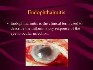
Endophthalmitis. Endophthalmitis is the clinical term used to describe the inflammatory response of the eye to ocular infection. Drugs 1996, 52(4), 526-540. Classification. Endophthalmitis can be classified according to the. Mode of entry Type of etiological agent Location in the eye.

Endophthalmitis
E N D
Presentation Transcript
Endophthalmitis • Endophthalmitis is the clinical term used to describe the inflammatory response of the eye to ocular infection. Drugs 1996, 52(4), 526-540
Classification Endophthalmitis can be classified according to the • Mode of entry • Type of etiological agent • Location in the eye
Exogenous endophthalmitisExogenous endophthalmitisis mostoften a postsurgical complication. It can also result from foreign-body penetration. Staphylococcus epidermidisandBacillus cereusare the most common bacterial agents involved. This child presented with a high fever and eye swelling, which was initially treated as conjunctivitis. When his eyelids were everted, the right globe was proptotic and the cornea was a cloudy white. An ophthalmologist diagnosed this as a panophthalmitis; enucleation was necessary.
EndophthalmitisPost operative, Bleb associted, Traumatic • Post-operative endophthalmitis is the most common form. • It comprises 70% of infective endophthalmitis
Postoperative endophthalmitis • May occur after any surgical procedure. • Possibility must be considered after any surgical procedure that breaches the integrity of the corneo-scleral wall of the eye, no matter how ‘minor’ the breach may be Ophthalmology 1998; 105(6): 1004-1010
Large majority follow cataract surgery, most common surgical procedure (approx prevalence 0.082%- 0.1%) • Post- operative endophthalmitis is one of the most dreadedcomplications of cataract surgery and constitutes a true emergency.
Incidence of postoperative endophthalmitis • Worldwide, the reported incidence of post-op endophthalmitis is 0.04-4%. Post cataract surgery 0.265% ( more with clear corneal incision) Post keratoplasty 0.382% Post Vitrectomy 0.05%. Bleb associated 0.2%-9.6% Post traumatic 2.4%-8%, retained IOFB 30%
POE: A potentially blinding condition • Though rare, it is potentially the most devastating complication of intraocular procedures and can lead to a permanent, complete loss of vision. (animal studies confirm that the retina begins to necrose very quickly in endophthalmitis) • Endophthalmitis has been associated with severe visual loss in 20% of patients. Surv Ophthalmol 2004, 49(2), S53-S54)
Post-op endophthalmitis: causes • Periocular flora gain access into eye during surgery • Organisms may be carried into the eye as surface fluid refluxes through the wound during surgery • IOL contamination if it touches the ocular surface or with the air of the operating room • Contaminated irrigation solutions
Risk factors Bacterial • Defects in sterilization of instruments. • Contamination of fluids and drugs • Complicated surgery (ruptre of posterior capsul),tissue damage • Lacrimal drainage obstruction Fungal • Contaminated irrigating solutions. • Contaminated IOLs, viscoelastics, poor OT hygiene, hospital construction activity.
Symptoms Patient presents with symptoms most commonly on the second day after surgery • Pain • Red eye • Decreased vision • Hazy cornea • Hypopyon
POE: Clinical aspects • Three forms of clinical presentation can be distinguished • Acute form, usually fulminant, occurs 2-4 days post-op, most commonly due to S.aureus or streptococci. • Delayed form, moderately severe, occurs 5-7 days post-op, due to S.epidermidis, coagulase negative cocci, rarely fungal. • Chronic form, occurs as early as 1 month post-op, due to Propionibacterium acnes, S.epidermidis or fungal.
Day of presentation of infection In most cases, infection occurs in immediate post-op period,
POE: Aetiological agents • Most common potential source of infection is the periocular flora of the patient • 75% of conjunctival cultures from normal eyes harbour Staph. epidermidis, Staph. aureus and various streptococci • Similar pattern has been found in eyes with post-operative endophthalmitis • Role of external ocular bacterial flora in the pathogenesis of post-op endophthalmitis has been proven by DNA studies
Coagulase-negative staphylococcal endophthalmitisafter intravitreal injection of ganciclovir for the treatment of cytomegalovirus retinitis. A hypopyon is present in the anterior segment. The view of the fundus was poor because of dense vitreitis. Treatment with intravitreal injection of antibiotics and vitrectomy controlled the infection, but vision was lost because of retinal detachment. A similar presentation with hypopyon could occur in acute endogenous endophthalmitis or in drug-induced uveitis
Pneumococcal endophthalmitisA, A 63-year-old man was admitted for acute blindness in the left eye. Three weeks before admission, he developed fever, productive cough with rusty sputum, and pleuritic chest pain. On admission, the patient had left lower lobe pneumonia, mitral valve endocarditis, meningitis, and endophthalmitis secondary to a septic embolus. Cultures of blood, fluid from the anterior chamber of the left eye, and spinal fluid were positive for type-8Streptococcus pneumoniae. B, Two weeks later, the endophthalmitis had worsened, necessitating eventual enucleation • .
There is a chronic exposure of intraocular contents to the tear film, and in some cases, endophthalmitis can develop years after the original surgery.
Blebitis • Blebitis. This condition, a microbial bacterial infection of the bleb without vitreous involvement, may complicate the postoperative course months to years after filtering surgery [ref], [ref]. The mnemonicRSVPis a useful reminder to patients and physicians of the warning signs and symptoms of blebitis and early endophthalmitis. The development ofR, redness (conjunctival injection or ciliary flush); S, sensitivity to light (photophobia); V, vision change (decreased central visual acuity); orP, pain (ciliary body spasm) in a patient who has had trabeculectomy demands immediate examination. All patients who have thin-walled blebs with or without microscopically observable leaks should be informed of the risks for late-onset bleb infections. The medical records of patients at risk for this complication should be identified to expedite emergency management.
Propionibacterium acnes endophthalmitisThis patient had cataract surgery 1 year before
Diagnosis • Clinical picture can be confirmed by culture of the organisms • The most important samples to culture are aspirates from the aqueous and vitreous cavity • Possibility of isolating an organism from vitreous 56-70% while from aqueous 36-40% www.aios.org
Obtaining aqueous samples • Aqueous fluid is obtained by paracentesis • About 0.1 ml fluid is aspirated • Innoculated on culture media www.aios.org
Obtaining vitreous samples • Sample of vitreous is a very important source to know the causative organisms • Aspiration may not provide adequate sample as vitreous is denser and contain inflammatory membranes in endophthalmitis • There is also chance of retinal detachment. • Safest method is vitreous biopsy (0.2-0.3 ml) • Lost volume of vitreous replaced by saline www.aios.com
Differential diagnosis:Other types of intraocular inflamationPreexisting Uveitis, Keratitis, Glaucoma therapy, Previous surgeryPseudohypopyon may be simulated by RBC, Debries, PigmentsRetained lens material cause sterile post op inflamationToxic anterior segment syndrome (TASS):causes hypopyon without infectionTumor cellsCare for inoculation due to unnecessary paracentesisProgressive vitritisout of proportion to other anterior segment findings = EndophthalmitisWhen doubt manage as infection
Delayed-onset endophthalmitis caused by Propionibacterium acnes
Management Findings of the Endophthalmitis Vitrectomy Study (EVS) provide guidelines for management of POE.
ENDOPHTHALMITIS VITRECTOMY STUDY Multicenter randomized trial carried out at 24 centres in U.S. (1990-1994) Purpose : To determine • The role of IV antibiotics in the management of POE • Role of initial vitrectomy in management. • Patients : N = 420 patients having clinical evidence of POE within 6 weeks of cataract surgery
EVS Intervention Random assignment toimmediate vitrectomy (VIT) or vitreous biopsy (TAP). They were also randomly assigned to treatment with IVor no IV. Medications :After initial VIT or TAP, all patients received intravitreal injection of amikacin (0.4 mg) + vancomycin (1 mg). Vancomycin (25 mg in 0.5 ml), ceftazidime (100 mg in 0.5 ml), dexamethasone (6 mg in 0.25 ml) were administered subconjunctivally. IV treatment: ceftazidime (2 g every 8 hrs) + amikacin (6mg/kg every 12 hrs) for 5-10 days Main outcome measure Evaluation of visual acuity and clarity of ocular media at 3, 9, 12 months
Results of EVS • Systemic antibiotics were of no benefit in this study. • Initial Vitrectomy was only beneficial for patients presenting with a very poor visual acuity.
Management • In established endophthalmitis, antibiotics when given oral or I.V. have poor penetration into the vitreous cavity. • Hence, intravitreal injections are treatment of choice. • Intravitreal injections rapidly achieves therapeutic levels at the sites of infection
For gram positive organisms • Because most cases are caused by gram positive organisms, vancomycin- (broad-spectrum activity against most gram positive species) has become an agent of choice • Thusvancomycin 1 mg in (0.1 ml) is given intravitreally • Non toxic in recommended clinical dosage. Arch Ophth 1999; 117: 1023-1027
Studies have proved that intravitreal vancomycin is the most effective drug for treating endophthalmitis • Administration of single intravitreal vancomycin dose results in adequate antibiotic concentrations for over one week
For gram negative organisms • Gentamicin (0.4 mg) was used, but was found to be associated with retinal toxicity • Amikacin was used (4 times less retinal toxicity than gentamicin as shown by animal studies) • Amikacin covers large number of gram negative organisms and those resistant to other aminoglycosides
A survey of retinal specialists suggested that amikacin can also cause retinal toxicity • Thus, Ceftazidine has emerged as on alternative to amikacin • More effective than aminoglycosides • Retinal toxicity studies in primates reveal concentration of 2.25 mg/0.1 ml to be safe.
Vancomycin combined with amikacin or ceftazidime appears to be best association in treatment of POE.
Steroids • Based on experimental studies in rabbits, an intravitreal injection of 0.2-0.4 mg of dexamethasone was recommended within first 10 hrs after inoculation (except when fungal infection is suspected) B J O 1997; 81: 1006-51
Prophylaxis • Pre-operative scrub • Povidone-iodine (5%) has broad antibacterial, as well as antifungal & antiviral activity • It decreases conjunctival flora growth to 91% • Can destroy bacteria in 30 secs
Role of prophylactic antibiotics Studies have shown that prophylactic antibiotic reduces the number of conjunctival bacteria at the time of surgery • Optimal choice of pre-operative topical antibiotic depends on spectrum of bacteria covered • Rapidity of killing • Duration of action • Penetration and toxicity of antibiotic • Antibiotic susceptibility pattern • Cost