Chapter 23
Chapter 23. The Respiratory System. Respiratory System Anatomy. Structurally , the respiratory system is divided into upper and lower divisions or tracts. The upper respiratory tract consists of the nose, pharynx and associated structures. The lower respiratory tract

Chapter 23
E N D
Presentation Transcript
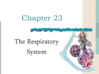
Chapter 23 The Respiratory System
Respiratory System Anatomy • Structurally, the respiratory system is divided into upper and lower divisions or tracts. • The upper respiratory tract consists of the nose, pharynx and associated structures. • The lower respiratory tract consists of the larynx, trachea, bronchi and lungs. Upper respiratory tract Lower respiratory tract
Respiratory System Anatomy • Functionally, the respiratory system is divided into the conducting zone and the respiratory zone. • The conducting zone is involved with bringing air to the site of external respiration and consists of the nose, pharynx, larynx, trachea, bronchi, bronchioles and terminal bronchioles. • The respiratory zone is the main site of gas exchange and consists of the respiratory bronchioles, alveolar ducts, alveolar sacs, and alveoli.
Respiratory System Anatomy • Air passing through the respiratory tract traverses the: • Nasal cavity • Pharynx • Larynx • Trachea • Primary (1o) bronchi • Secondary (2o) bronchi • Tertiary (3o) bronchi • Bronchioles • Alveoli (150 million/lung)
The Nose • The external nose is visible on the face. • It consists of: • a supporting bony frame- work (frontal bone, nasal bones, and maxillae) and a • cartilaginous framework of hyaline cartilage
The Nasal cavity • Lies in and posterior to the external nose • Is divided by a midline nasal septum • Formed by the perpedicular plate of ethmoid, & the vomer posteriorly and the septal cartilage anteriorly • It opens posteriorly into the naso- pharynx Wikimedia Commons
Nasal Cavity- lateral wall • Three nasal conchae (or turbinates) protrude medially from each lateral wallof nasal cavity • Superior concha • Middle concha • Inferior concha • Increase mucosal surface area & air turbulence- ensures air contacts mucosa • Under each nasal concha is an opening, or meatus, for a duct that drains secretions of the sinuses and tears into the nose.
The Nose • Functions: • Providing an airway for respiration • Moistening and warming & filtering inspired air • Resonation of sound • Olfaction
The Paranasal Sinuses • Mucosa-lined, air-filled spaces found in five skull bones – the frontal, sphenoid, ethmoid, and paired maxillary bones • Sinuses lighten the skull and help to warm and moisten the air
The Paranasal Sinuses Mucosal secretions flows from the sinuses into nasal cavity
The Phrynx • The pharynx is a hollow tube that starts posterior to the internal nares and descends to the opening of the larynx in the neck. • It is formed by a complex arrangement of skeletal muscles that assist in deglutition. • It functions as: • a passageway for air and food • a resonating chamber • a housing for the tonsils
The Pharynx • The pharynx has 3 regions • The nasopharynx is separated from the oropharynx by the hard and soft palate Nasopharynx Oropharynx Laryngopharynx
The Nasopharynx • Lies posterior to the nasal cavity and superior to the level of the soft palate • Strictly an air passage • Lined with psuedostratified columnar epithelium • Closes during swallowing to prevent food from entering the nasal cavity • The pharyngeal tonsil ( adenoids) lies high on the posterior wall • Auditory tubes from middle ears open into the lateral walls
Respiratory Lining • Cilia in the upper respiratory tract move mucous and trapped particles down toward the pharynx. • (Cilia in the lower respiratory tract move them up toward the larynx.)
The Pharynx • The oropharynx & laryngopharynx are both common passages for food and air & are lined by stratified squamous epithelium • The oropharynx lies posterior to the oral cavity & opens into the oral cavity via the fauces • The palatine tonsils lie in the lateral walls of the fauces (those usually taken in a tonsillectomy) and small lingual tonsil at the base of the tongue • The laryngopharynx lies posterior to the upright epiglottis • Leads into the larynx & the esophagus
The Larynx • The larynx, composed of 9 pieces of cartilage, forms a short passageway connecting the laryngopharynx with the trachea (the “windpipe”). • The thyroid cartilage (the large “Adam’s apple”) and the one below it (the cricoid cartilage) are landmarks for making an emergency airway (called a cricothyrotomy). Anterior view of the larynx
The Larynx • 9 Cartilages of the larynx • Epiglottis – elastic cartilage that covers the glottis during swallowing • Thyroid cartilage- hyaline cartilage with a midline laryngeal prominence (Adam’s apple) • Cricoid cartilage - hyaline cartilage • Three pairs of small arytenoid,corniculate, & cuneiform cartilages
The Larynx • The epiglottis is a flap of elastic cartilage covered with a mucus membrane, attached to the root of the tongue. • The epiglottis guards the entrance of the glottis, the opening between the vocal folds. • For breathing, it is held anteriorly, then pulled back- ward to close off the glottic opening during swallowing.
The Larynx • The mucous membrane of the larynx forms two pairs of folds: • The superior pair are the Ventricular folds ( false vocal cords) -also called vestibular folds • The space between the ventricular folds is the rima vestibuli • The inferior pair are the vocal folds ( true vocal cords) • The space between the vocal folds ( true vocal cords) is the rima glottidis • True vocal cords & the opening between them form the glottis
The Larynx • The functions of the larynx are: • To provide an airway • To route air and food into the proper channels • To function in voice production- True vocal cords vibrate to produce sound as air passes • False vocal cords have no part in sound production; help close glottis during swallowing • Valsalva’s maneuver- by closing the glottis the larynx is closed during certain abdominal straining conditions to prevent exhalation
Lower Respiratory Tract • As air passes from the laryngopharynx into the larynx, it leaves the upper respiratory tract and enters the lower respiratory tract. • Air passing through the respiratory tract • Nasal cavity • Pharynx • Larynx • Trachea • Primary bronchi • Secondary bronchi • Tertiary bronchi • Bronchioles • Alveoli (150 million/lung) Upper respiratory tract Lower respiratory tract
The Trachea • The trachea is a semi-rigid pipe made of semi-circular cartilaginous rings, and located anterior to the esophagus. • It is about 12 cm long and extends inferior to larynx into the mediastinum • At the level of carina ( an internal ridge of last tracheal cartiage) it divides into right and left primary (1o, “mainstem”) bronchi. • It is composed of 4 layers: the mucosa ( lined by ciliated respiratory epithelium), submucosa, hyaline cartilage, andadventitia
The Trachea • The tracheal cartilage rings are incomplete posteriorly, facing the esophagus. • Esophageal masses can press into this soft part of the trachea and make it difficult to breath, or even totally obstruct the airway.
The Bronchi • The right and left primary (1o or “mainstem”) bronchi emerge from the inferior trachea to go to the lungs • Right primary bronchus is more vertical compared toleft primary bronchus
The Bronchi • Primary bronchi- subdivide into: • Secondary bronchi (lobar bronchi),each supplying alobe of the lungs –two on the left side and three on the right • Subdivide intotertiary bronchi (segmental bronchi)- each supplies one bronchopulmonary segment • There are upto 10 bronchopulmonary segments in each lung http://pblnotes.wordpress.com/2011/
Bronchioles • Air passages undergo 23 orders of branchings • Bronchioles- smaller than 1mm in diameter- lack cartilage • Bronchioles divide into terminal bronchioles • A branch of the terminal bronchioles supplies air to a lobule • Terminal bronchioles branch into respiratory bronchioleswhich now have alveoli • Respiratory bronchioles lead to the alveolar ducts which have alveoli • The respiratory bronchioles, alveolar ducts and alveoli form the 'respiratory zone'
Lung lobule • Pulmonary lobule: • Wrapped in elastic C.T., each pulmonary lobule contains a lymphatic vessel, an arteriole, a venule and a branch of terminal bronchiole.
The bronchi and bronchioles go through structural changes as they branch and become smaller. • The mucous membrane changes • The cartilaginous rings become more sparse, and eventually disappear altogether. • As cartilage decreases, smooth muscle (under the control of the Autonomic Nervous System) increases. • Sympathetic stimulation causes airway dilation, while parasympathetic stimulation causes airway constriction.
All the branches from the trachea to the terminal bronchioles are conducting airways – they do not participate in gas exchange.
Alveoli • Alveoli are the cup-shaped outpouchings which participate in gas exchange • Alveoli make up a large surface area (750 ft2). • They are lined chiefly by type I alveolar cells, simple squamous epithelium)which allow for exchange of gases with the pulmonary capillaries.
Alveoli • Type II cells in the alveoli secrete a substance called surfactant that prevents collapse of the alveoli • Alveoli macrophages (also called “dust cells”) engulf and remove pathogens & debris
Respiratory Membrane • The Respiratory membrane across which diffusion of gases occurs is composed of: • Alveolar lining epithelium • Capillary endothelium • Their fused basement membranes
Blood Supply to the Lungs The lungs receive blood via two sets of arteries Pulmonary arteriescarry deoxygenated blood from the right heart to the lungs for oxygenation Bronchial arteries branch from the aorta and deliver oxygenated blood to the lungs primarily perfusing the muscular walls of the bronchi and bronchioles ( not the alveoli)
The Lungs • The lungs are divided into lobes by fissures. • The right lung is divided by the oblique fissure and the horizontal fissure into 3 lobes . • The left lung is divided into 2 lobes by the oblique fissure. • Each lobe receives it own 2o bronchus that branches into 3osegmental bronchi (which continue to further divide).
Respiratory System Anatomy • The apex of the lung is superior, and extends slightly above the clavicles. The base of the lungs rests on the diaphragm. • The cardiac notch – in the left lung (the indentation for the heart) • The medial mediastinal surface has the hilus– an indentation
Respiratory System Anatomy • The lungs are separated from each other by the heart and other structures in the mediastinum. • Each lung is enclosed by a double-layered pleural membrane. • The parietal pleura line the walls of the thoracic cavity. • The visceral pleura adhere tightly to the surface of the lungs themselves.
Respiratory System Anatomy • On each side of the thorax, a pleural cavity is formed. • The pleural cavity contains pleural fluid -reduces friction • The pleura, adherent to the chest wall and to the lung, produces a mechanical coupling for the two layers to move together.