Download
1 / 23
280 likes | 636 Views
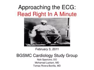
Approaching the ECG: Read Right In A Minute. BGSMC Cardiology Study Group Nick Sparicino, DO Mohamad Lazkani, MD Tomas Rivera-Bonilla, MD. February 3, 2011. History of the ECG/EKG.

E N D
Approaching the ECG:Read Right In A Minute BGSMC Cardiology Study Group Nick Sparicino, DO Mohamad Lazkani, MD Tomas Rivera-Bonilla, MD February 3, 2011
History of the ECG/EKG • During the late 1800’s and early 1900’s, Dutch physiologist Willem Einthoven developed the early electrocardiogram He won the Nobel prize. • Hubert Mann first uses the electrocardiogram to describe electrographic changes associated with a heart attack in 1920 • Electrocardiograms must be viewed in the context of demographics, health history, and other clinical test correlates. They are especially useful when compared across time to see how electrical activity of the heart has changed (perhaps as the result of some pathology). • Why PQRST and not ABCDE? The choice of P is a mathematical convention dating from Descartes by using letters from the second half of the alphabet. N has other meanings in mathematics and O is used for the origin of the Cartesian coordinates. P is simply the next letter. (For more on Descartes see Henson JR. Descartes and the ECG lettering series. J Hist Med Allied Sci. April 1971;181�186)
Electrocardiography • A recording of the electrical activity of the heart over time • Gold standard for diagnosis of cardiac arrhythmias • Helps detect electrolyte disturbances (hyper- & hypokalemia), arrhythmias, myocardial ischemia and infarction, pericarditis, chamber hypertrophy, drug toxicity (i.e. digoxin and drugs which prolong the QT interval) • Allows for detection of conduction abnormalities • Screening tool for ischemic heart disease during stress tests • Helpful with non-cardiac diseases (e.g. pulmonary embolism or hypothermia)
Electrocardiogram (ECG/EKG) • Is a recording of electrical activity of heart conducted through ions in body to surface
ECG Rules • Wave of depolarization traveling towards a positive electrode causes an upward deflection on the ECG. • Wave of depolarization traveling away from a positive electrode causes a downward deflection on the ECG.
Vector 1 – depolarization of atrium(corresponds to P wave) Vector 2 – Ventricular Septum (1st deflection of QRS) Vector 3 – Bulk of ventricular muscle Vector 4 – Repolarization of ventricular muscle Vectors: directions and amplitude
Approach to the ECG • Systematic Approach • rhythm, rate, intervals, axis, morphology RRIAM: Read Right In AMinute
RHYTHM • Locate the P wave • Establish the relationship between P waves and QRS complex • If no P wave analyze the QRS morphology • Search for other clues • Interpret the rhythm in the clinical setting
RATE • Determining rate: • Regular rhythm: • Big box: 300, 150, 100, 75, 60, 50 • Irregular rhythm: • # cycles in a 6 second strip x10 • # cycles in a 12 second strip x5 • remember to use halves if half a cycle is present in the strip 10mm = 1mV 1mm = 0.1mV
RATE: Intrinsic rates of pacing cells (transmits the impulses through the inter-atrial septum) • SA node 60-100 BPM • Atrial cells 55-60 BPM • AV node 45-50 BPM • His bundle 40-45 BPM • Bundle branch 40-45 BPM • Purjkinje cells 35-40 BPM • Myocardial cells 30-35 BPM
ATRIAL COMPONENTS • P wave – atrial depolarization • Duration 0.08 to 0.12 sec • PR interval - impulse initiation, atrial depol, atrial repol, AV/His/BB/Purkinje stimulation • 0.12 to 0.20 seconds (>0.20 seconds = PR prolongation; Heart Block discussed later)
VENTRICULAR COMPONENTS • QRS complex - ventricular depolarization • 0.06 to 0.12 seconds • Q wave Significance • “pathological/MI” >0.03s or >1/3 height of R wave
VENTRICULAR COMPONENTS QT Interval - all the events of ventricular systole • Beginning of QRS to end of T wave • Duration varies with heart rate, age, sex but should be less than half the RR interval • Correction formulas exist to balance HR, a major variable (as HR decreases, QT interval increases) • Fridericia Correction (QTf): • QTf = QT interval / cubed root of the RR interval (in sec) • Bazett’s formula (QTc): • QTc = QT interval / square root of the RR interval (in sec)
VENTRICULAR COMPONENTS • ST segment - electrically neutral period between ventricular depol and repol (time myocardium is maintaining contraction in order to push the blood out of the ventricles) • T wave - ventricular repolarization • Should be asymmetrical with a slow upstroke and a fast downstroke
AXIS - Quadrant Graphing Method I (+) aVF (+) =normal I (+) aVF (-) =LAD I (-) aVF (+) =RAD I (-) aVF (-) =extreme LAD or RAD
AXIS - Isoelectric Method 1 2 • Find isoelectric lead • Find perpendicular lead • If QRS positive, vector towards lead, if negative, away
MORPHOLOGY • Hypertrophy (atrial & ventricular) • Bundle branch blocks and hemiblocks • Segment depressions & elevations • PR segment, ST segment • U waves • T wave morphologies • Delta wave • Osborne wave • ST – T changes
Hypertrophy Criteria • Left atrial enlargement • Terminal negative P wave deflection in V1 > 0.04s and the amplitude of “same” P wave in V1 > 0.10mV • P-mitrale in lead II (notched P wave) Duration b/w peaks of P wave notches >0.04s • Max p wave duration >0.11s • Ratio of P wave duration to PR duration > 1:1.6 • Right atrial enlargement • P wave amplitude >2.5 mm in II and/or >1.5 mm in V1 (these criteria are not very specific or sensitive) • Better criteria can be derived from the QRS complex; these QRS changes are due to both the high incidence of RVH when RAE is present, and the RV displacement by an enlarged right atrium. • QR, Qr, qR, or qRs morphology in lead V1 (in absence of coronary heart disease) • QRS voltage in V1 is <5 mm and V2/V1 voltage ratio is >6 • Biatrial enlargement • Features of both RAE and LAE in same ECG • P wave in lead II >2.5 mm tall and>0.12s in duration • Initial positive component of P wave in V1 >1.5 mm tall and prominent P-terminal force
Hypertrophy Criteria • Other LVH Criteria • Sokolow-Lyon Criteria • S wave in V1 + R in V5 or V6 >= 3.5mV • Or R wave in V5 or V6 >2.60mV • Cornell Voltage Criteria • Female: R in aVL + S in V3 >2.0 mV • Male: R in aVL + S in V3 > 2.8 mV • Hypertrophic Cardiomyopathy • LVH (tall R in V2-V5) • Deep narrow Q in aVL and V6 • LAE (increased negative terminal p wave in V1) • Other Criteria (quick glance) • I = R >14mm • aVR = S >15 mm • aVL = R > 12 mm • aVF = R >21 mm • V5 = R > 26 mm • V6 = R >20 mm
Hypertrophy Criteria RVH Criteria • Right ventricular hypertrophy • Any one or more of the following (if QRS duration <0.12 sec): • Right axis deviation (>90 degrees) in presence of disease capable of causing RVH • R in aVR > 5 mm, or • R in aVR > Q in aVR • Any one of the following in lead V1: • R/S ratio > 1 and negative T wave • qR pattern • R > 6 mm, or S < 2mm, or rSR' with R' >10 mm • Other chest lead criteria: • R in V1 + S in V5 (or V6) 10 mm • R/S ratio in V5 or V6 < 1 • R in V5 or V6 < 5 mm • S in V5 or V6 > 7 mm • ST segment depression and T wave inversion in right precordial leads is usually seen in severe RVH such as in pulmonary stenosis and pulmonary hypertension.
Hypertrophy Criteria • Biventricular Criteria • 1. High voltage, biphasic RS complex in midprecordial leads (also common in LV septal defect) • 2. LVH criteria in precordial leads with RAD in limb leads • 3. Low amplitude S in lead V1 combined with deep S wave in lead V2 • 4. LVH criteria in left precordial leads combined with prominent R waves in right precordial leads • 5. LAE as sole criteria for LVH combined with any criteria suggestive of RVH • Left or right strain pattern Criteria • ST-T wave changes associated with abnormal repolarisation secondary to increased ventricular tension have classically referred to as "strain" pattern. • Left ventricular hypertrophy is often associated with ST depression and deep T wave inversion. These changes occur in the left precordial leads, V5 and V6. In the limb leads the ST-T changes occur opposite the main QRS forces. Therefore, if the axis is vertical, the ST-T changes are seen in II, III and aVF. If the axis is horizontal the ST-T changes are seen in I and aVL. • Right ventricular hypertrophy can be associated with ST depression and T wave inversion in the right precordial leads, V1 - V3. Leads II, II and aVF may also show similar ST - T wave changes.
Put it all together • Try to come up with a common theme to the differential diagnosis based on the list of abnormalities you’ve created • Do not overlook anything • Practice, Practice, Practice (look at many ECGs, especially normal ones in the beginning, developing good habits using RRIAM and calculate intervals committing normal values to memory)