Download
1 / 10
100 likes | 325 Views
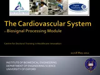
Institute of Biomedical Engineering Department of Engineering Science University of Oxford. Heart Sounds in Biosignal Processing Module Centre for Doctoral Training in Healthcare Innovation 11/18 May 2012. S1 – Atrial contraction S2 – Ventricular contraction

E N D
Institute of Biomedical Engineering Department of Engineering Science University of Oxford Heart Soundsin Biosignal Processing ModuleCentre for Doctoral Training in Healthcare Innovation11/18 May 2012
S1 – Atrial contraction S2 – Ventricular contraction S3 – Blood returning to the ventricle S4 – Ventricle is too full to contain the blood How a heart sounds Animation of heart valves opening and closing with sound “LUB” “DUB” Normal Abnormal S1 + S2 Lub - Dub S1 + S2 + S3 Ken – Tuck – Y S4 + S1 + S2 Ten – Nes - See
Heart sounds http://commons.wikimedia.org/wiki/File:Phonocardiograms_from_normal_and_abnormal_heart_sounds_with_pressure_diagrams.png How heart sounds change during illness
Heart sounds Chest piece transmits sound to the listener via air-filled hollow tubes • How to record heart sounds? • Acoustic Stethoscope
Heart sounds Place a microphone in the chestpiece Moving magnetic coil electrical signal • How to record heart sounds? • Electronic Stethoscope - microphone
Heart sounds Connect one end of the crystal with the diaphragm Plates squeezed electric signal • How to record heart sounds? • Electronic Stethoscope - piezoelectric
Heart sounds Distance between plates Body Electromagnetic diaphragm with conductive inner surface Vibrations electric signal Conductive plates • How to record heart sounds? • Electronic Stethoscope - capacitor
Heart sounds Ascultation – library of heart sounds Plug-in stethoscope iStethoscope – turn your iPhone into a stethoscope Phone apps
Heart sounds Other uses of stethoscopes:
Heart sounds Any Questions?