Download
1 / 83
1.32k likes | 5.1k Views
SYNCOPE EVALUATION AND MANAGEMENT. Jayne Barr, MD Clinical Assistant Professor The Ohio State University. Case #1. 42 year old female Chief complaint: passing out at work Works in a pharmaceutical lab. Was sitting at her desk, felt nauseated and knew she was going to pass out.

E N D
SYNCOPEEVALUATION AND MANAGEMENT Jayne Barr, MD Clinical Assistant Professor The Ohio State University
Case #1 • 42 year old female • Chief complaint: passing out at work • Works in a pharmaceutical lab. Was sitting at her desk, felt nauseated and knew she was going to pass out. • Per witnesses, was slump over chair and was unconscious for a few seconds.
Case continues • No chest pain, palpitations, shortness of breath • Similar episodes 10 years ago. • No family history of sudden cardiac death • No medications. • No smoking. No alcohol. No drugs.
Case #2 • 82 year old male. • Found unresponsive by his son • Past medical history—HTN • Medications—HCTZ • Exam—BP 160/98. P 70. Now alert and oriented. Facial contusions. Otherwise normal exam. Q: How would you manage these 2 patients?
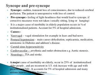
Syncope: Definition • Abrupt and self-limited loss of consciousness associated with absence of postural tone • Relatively rapid onset. Variable warning symptoms. • Followed by rapid and complete recovery. Last only a few minutes. • Absence of prolonged confusion • Presyncope---prodromal symptom of fainting and typically has the same work up as syncope.
Significance of Syncope • “The only difference between syncope and sudden death is that in one you wake up”. [1] • --anonomymous
Syncope: Epidemiology • 20-50% of adults experience at least one episode of syncope during their lifetime. • Explained 53-62% • Infrequent, unexplained 38-47% • 500,000 new syncope patients each year. • 3-5% of ER visits. • 6% of hospital visits. • More common in the elderly. • Up to 23% in age >70 years.
Per recent data, the overall cost per hospital admission was estimated to be about $10,600. One study found to be $17,000 of “unnecessary” testing to diagnosis vasovagal syncope Overall cost in US estimated to be in excess of $1 billion. Costs of Test Troponin $78 EKG $221 Telemetry $255/d Head CT $1545 MRI brain $2216 Carotid US $1294 EST $2492 Echocardiogram $809 EEG $1115 Syncope: Economic Burden
Morbidity and Mortality • Most cases benign. • Syncope of cardiac origin has the highest morbidity and mortality. • 1 year mortality of 18-33% • Recurrence in the elderly population is 30% • Syncope of unknown origin. • 1 year mortality of 6-12%.
Generally a benign event. Most common causes Vasovagal (40%) Simple faint (29%) Breathholding (4%) Unknown (15%) Rare but serious causes of syncope in children Hypertrophic cardiomyopathy Anomalous origin of left coronary artery Myocarditis Long QT syndrome Cystic medial necrosis WPW Syncope in Children
Syncope: Pathophysiology • Decreased cerebral perfusion is common to all causes of syncope • Cessation of cerebral perfusion for as little as 3-5 seconds can result in syncope • Decreased cerebral perfusion may occur as a result of decreased cardiac output or decreased systemic vascular resistance.
Bilateral hemisphere dysfunction or reticular activating system (RAS) midbrain knockout. Generally acute hypoperfusion is responsible. Regional (vasoconstriction) Systemic (global hypotension) Loss of consciousness causes loss of postural tone leading to collapse 35% reduction in cerebral blood flow will cause syncope. Modifying factors Cardiac output Systemic and local vascular resistance/occlusion Blood volume Ability to compensate More Pathophysiology
Syncope: Etiology • Mnemonic: PASSOUT • P-ressue (hypotensive causes) • A-rrthymias • S-eizures • S-ugar (hypo/hyper glycemia) • O-utput (cardiac)/ O2 (hypoxia) • U-nusual causes • T-ransient (TIAs, strokes, CNS diseases)
OUTPUT Cardiac AS, PA, MS,IHSS Cardiomyopathies Atrial myoxomas Cardiac tamponade Aortic dissection MI, CHF Pulmonary PE, acute hypoxemia Pulmonary HTN COPD exacerbation CO poisoning UNUSUAL CAUSES Anxiety, Panic disorder Major depressive disorder Somatization disorder (psychogenic syncope) Hyperventilation syndrome Migraine, sleep disorder TRANSIENT TIA (vertebrobasilar), CVA, subdural hematoma Subarachnoid hemorrhage CNS mass effect (tumor) Basilar artery migraine. More specifics
Syncope • CAUSES (Head---Heart---Vessels) • Reflex mediated • Vasovagal, carotid sinus, situational • Cardiac • Mechanical , arrhythmias • Orthostatic • Drugs, autonomic failure • Cerebrovascular • Unknown • Nonsyncopal causes
Neurally mediated reflex syncope(36-62%); average (24%) • Vasovagal, carotid sinus, situational • No increased risk for cardiovascular morbidity or mortality associated with reflex mediated syncope.
Neurally Mediated Reflex Syncope--what happens? • Stress causes an abnormal autonomic reflex • Normal increased sympathetic tone replaced by increased vagal tone • Variable contribution of vasodilation and bradycardia. • Examples include syncope from: • Pain and/or fear • Carotid sinus hypersensitivity • “situational” (cough, micturition, defecation syncope)
Vasovagal syncope • Most common cause of syncope in young adults • Precipitating event is often identifiable • Stress, trauma, pain, sight of blood, prolonged standing, heat exposure
Vasovagal Syncope • 3 PHASES • --Prodrome • Diaphoresis, epigastric discomfort, weakness, nausea, dizziness • Lasts about 2 minutes • --Loss of consciousness • Usually lasts 5-20 seconds • --Postsyncopal phase • Nausea, dizziness, general sense of poor health • If present, confusion which lasts no more than 30 seconds
Prevalence of VasoVagal Syncope • Prevalence poorly known (8-37% with mean of 18%) • Important points • Patients with VVS younger than Carotid sinus syndrome patients • Age range teens to elderly with mean 43 years • Pallor, nausea, sweating, palpitations are common • Amnesia for warning symptoms in older patients
Management for Vasovagal syncope • Optimal management is source of debate • Patient education, reassurance, instruction • Fluids (sports drinks), salt, diet • Tilt training • Support hose (waist high) • Drug therapies • Pacing (DDD pacing) • Class II indication if positive tilt test and cardioinhibitory or mixed reflex
Salt/volume Salt, sports drinks, fludrocortisone Beta-adrenergic blockers 1 positive control study using atenolol Use if hx of htn Disopyramide SSRIs 1 controlled study Use if hx of depression Vasoconstrictors (eg, midodrine) 1 negative controlled study (etilephrine) ? Efficacy of neosynephrine Use midodrine if significant hypotension Drug therapies for Vasovagal syncope
Postural Orthostatic Tachycardia Syndrome • Upright symptoms without hypotension. • Upright tachycardia—excessive HR response to maintain a low normal BP. • 500,000 Americans, usually young women • Partial dysautonomia • Antecedent infection, surgery, pregnancy • Treatment—low dose propanolol 10mg tid
Carotid Sinus Syncope • Syncope related to head turning, shaving, wearing a tight collar • Pathophysiology • Carotid sinus pressure causes a reflex decrease in heart rate and blood pressure
Site Carotid arterial pulse just below thyroid cartilage Method Massage, not occlusion. Right followed by left, pause between Duration:5-10 seconds Posture: supine and erect Risks 1/5000 massages complicated by TIA Outcome 3 sec asystole and/or 50mmHg fall in systolic blood pressure with reproduction of symptoms ==CAROTID SINUS SYNDROME Contraindications Carotid bruit, known but significant carotid arterial disease, previous CVA, MI last 3 months. Carotid sinus massage
Situational Syncope • Related to micturition, defecation, swallowing or coughing • Induced by baroreceptor and mechanoreceptors causing vagal stimulation • Circumstances of the event are typically diagnostic
Orthostatic syncope • When vertical, blood follows gravity and pools. • Increased sympathetic tone counteracts this. • If the response is inadequate, syncope occurs. • Drop in BP: 20 systolic or 10 diastolic within 3 minutes of standing • Present in 40% of patients over 70 years old • May be due to • Drugs • Volume loss • Neurologic damage
Volume loss Assoc. with tachycardia Medications Seen in elderly 45% of time Situational Micturition, cough, postprandial, carotid sinus sensitivity, defecation, laughing Adrenal insufficiency Primary autonomic disease Idiopathic, parkinsons disease, multisystem atrophy (Shy-Dragger) Secondary autonomic disease Neuropathic (dm, amyloid, alcoholism, autoimmune, vitamin deficiency, etc) CNS (cva, MS, tumors, spinal cord) More on Orthostatic Hypotension
Two basic types Dysrhythmia mediated Structural cardiopulmonary lesions Both cause the heart to be unable to sufficiently increase cardiac output to meet demand Double the risk of mortality compared with other syncopal patients. Up to 50% mortality. Patients with underlying cardiac disease are at greatest risk for cardiac syncope. Only 3% have no previous heart disease. Cardiac arrythymias especially in the elderly have high mortality. Cardiac Syncope
Neurologic Syncope • Rarely the primary cause of syncope • Ischemia to the RAS in the brainstem may cause “drop attacks” • Results from Vertebrobasilar insufficiency due to TIA (sometimes basilar migraine) • Usually accompanied by vertigo, ataxia, dysarthia, diplopia • Other examples • Subclavian steal—occurs with arm activity. Systolic BP in arms (difference of 10mmHg) • Subarachnoid hemorrhage
Psychiatric causes • Most commonly associated with • Anxiety • Panic • Major depressive disorders • Variety of mechanisms may be involved • Hyperventilation • Increased vagal tone
Syncope-like States • Migraine • Acute hypoxia • Hyperventilation • Somatization disorder (psychogenic syncope) • Acute intoxication (ie alcohol) • Seizures • Hypoglycemia • Sleep disorders
HISTORY • RAPID ASSESSMENT • Identify Life-Threatening causes • Dysrhythmias • cardiac ischemia • Critical aortic stenosis • Aortic dissection • Pulmonary embolus • CVA • SAH • Toxic-metabolic derangement
HISTORY • HISTORY alone identifies the cause up to 85% of the time • POINTS • Previous episodes • Character of the events, witnesses • Events preceding the syncope • Events during and after the episode
Events preceding the syncope Prolonged standing (vasovagal) Immediately upon standing (orthostatic) With exertion (cardiac) Sudden without warning or palpitations (cardiac) Aggressive dieting Heat exposure Emotional stress Events during and after the episode Trauma (implication important) Chest pain (CAD, PE) Seizure (incontinence, confusion, tongue laceration, postictal behavior) Cerebrovascular syndrome (diplopia, dysarthia, hemiparesis) Associated with n/v/sweating (vasovagal) HISTORY
Associated symptoms Chest pain, SOB, lightheadedness, incontinence Past medical history Identifying risk factors Morbidity and mortality increases with organic causes Parkinsons (orthostatic) Epilepsy (seizure) DM (cardiac, autonomic dysfunction, glucose) Cardiac disease Medications Antihypertensives, diuretics (orthostatic) Antiarrthymics (cardiac syncope) TCA, Amiodarone (cardiac/prolonged QT) Family history Sudden death (cardiac syncope/prolonged QT or Brugada) HISTORY
Vital signs Orthostatics—most important Drop in BP and fixed HR ->dysautonomia Drop in BP and increase HR -> volume depletion/ vasodilatation Insignificant drop in BP and marked increase in HR -> POTS Temperature Hypo/hyperthermia (sepsis, toxic-metabolic, exposure) Heart rate Tachy/brady, dysrhythmia Respiratory rate Tachypnea (pe, hypoxia, anxiety) Bradypnea (cns, toxicmetabolic) Blood pressure High (cns, toxic/metabolic) Low (hypovolemia, cardiogenic shock, sepsis) PHYSICAL EXAM
HEENT Tenderness/deformity (trauma) Papilledema (increased icp, head injury) Breath (alcohol, dka) NECK Bruits JVD (chf, mi, pe, tampnade) HEART Murmur (valves, dissection) Rub (pericarditis, tamponade) LUNGS Sounds may help distinguish chf, infection, pneumothorax PHYSICAL EXAM
ABDOMEN Pulsatile mass; AAA Tenderness Occult blood loss PELVIS Bleeding, hypovolemia Tenderness (PID, ectopic, torsion, sepsis) SKIN Signs of trauma, hypoperfusion EXTREMITES Paralysis (CNS) Pulses unequal (dissection, embolus, steal) PHYSICAL EXAM
NEUROLOGIC Mental status; toxic metabolic; organic disease; seizure; hypoxia. Focal findings (hemorrhagic/ischemic stroke, trauma, tumor, or other primary neurologic disease Cranial nerves Cerebellar testing PHYSICAL EXAM
SEIZURE Frothing at mouth Tongue biting Disorientation/ postictal Age < 45 year LOC over 5 minutes *tongue biting found only in seizure (99% specificity); absence did not exclude the possibility of a seizure (24% sensitivity) NOT A SEIZURE Sweating prior to episode Nausea prior to episode Oriented after event Age > 45 years Seizure or Not?
Ancillary Studies • EKG---Cornerstone of workup • Arrhythmia, long qt, WPW, conduction abn. • Routine Blood work—limited value • Radiology---limited value except if abnormal exam • Other tests—depending of history and exam • Glucose --hemoglobin --troponin • Ua/culture --CK (syncope vs seizure)
Starting the “Workup” • If young adult and No comorbid conditions or symptoms Most likely VASOMOTOR or ORTHOSTATIC . *Clinicians may forego the EKG in young, healthy patients with an obvious cause of syncope.
Normal EKG • If Normal EKG: • Check orthostatics • Check hemoglobin • If low---Anemia • If normal or high--Volume loss, dehydration, drug induced
Vasomotor Try carotid massage (+) carotid sinus sensitivity (-) reflex or neurocardiogenic Metabolic Check chemistry. R/O hypoglycemia, adrenal insufficiency Neurologic CT head (tia, cva, sah) EEG (if suspect Sz) Cardiovascular If Outflow obstruction, check CT chest, Echo (PE, valvular, HOCM) If venous return, check HCG, Echo (pregnancy, tamponade) Young adult, no comorbidity, normal EKG, absent orthostatics
The EKGKey Points • Guidelines recommend EKG in the evaluation of all patients with syncope. • Exception: young healthy patients with an obvious cause of syncope • Abnormal EKG in 90% of patient with cardiac syncope • Only 6% of patients with reflex mediated syncope have abnormal EKG. • Syncopal patient with negative cardiac history and normal EKG—unlikely to have a cardiac cause
The EKG patient older, +comorbid signs/symptoms • If Abnormal EKG • Ischemia/injury • Dysrhythmia • Sinus brady, BBB, AV block, prolonged QT, WPW, HOCM, Brugada • If Normal EKG • Consider holter or event recorder if dysrhythmia suspected