Download

1 / 36
360 likes | 375 Views
Explore the use of RNA interference (RNAi) to eliminate gene expression in human cells and its impact on phenotypic traits. Learn how to engineer gene deletions and perform genomic phenotyping using RNAi technology, fluorescent proteins, and DNA microarrays.

E N D
BEH.109: Laboratory Fundamentals in Biological Engineering. MODULE 3Eukaryotic Cells as Phenotypic Indicators:The use of RNAi to modulate gene expressionInstructor: Leona D. SamsonTeaching Assistants: Jenn Cheng and Lisa JoslinWith additional invaluable help from Lisa Smeester and Rebecca Fry
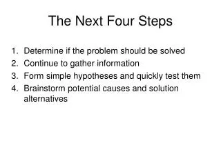
Snapshot of the next four weeks We will eliminate the expression of six different genes using RNAi technology, human cells, fluorescent proteins and DNA microarrays
The use of RNAi to modulate gene expression Why do we want to be able to modulate gene expression?
What is mRNA? GCU = Ala
“Genes” were first described by their mutant phenotype… e.g., Mendel described inherited properties like wrinkled versus smooth peas…later Bateson coined the word “gene” to account for these phenotypic traits. Genes were said be inherited in a Mendelian fashion.
1940’s Beadle and Tatum’s classic experiment with moulds established the “one gene one enzyme” hyothesis
In the 1940’s Beadle and Tatum mutated genes to analyze biochemical pathways Mutagens? X-rays, Nitrogen Mustards
1940’s Beadle and Tatum’s classic experiment with moulds established the “one gene one enzyme” hyothesis
Eliminating the expression of a gene is one of the most powerful tools in biology We can now engineer the deletion of specific genes to probe their biological funtion
Forward Genetics: Phenotype Genotype Reverse Genetics Genotype Phenotype
The most common method for Reverse Genetics has been Targeted Gene Deletion
Number of Genes in Different Organisms Human ~ 30,000 genes Mouse ~ 30,000 genes Yeast ~ 6200 genes E. coli ~ 4200 genes Phage T4 ~ 200 genes Influenza ~ 12 genes
1997 saw the sequencing of the first eukaryotic genome…. S. cerevisiae The field is changed for ever...
What does knowing the sequence of all 6,200 genes enable? Making 6,200 Gene Deletion Strains!
Genomic Phenotyping Using MMS WT mag1D rev1D rad14D
Number of Genes in Different Organisms Human ~ 30,000 genes Mouse ~ 30,000 genes Yeast ~ 6200 genes E. coli ~ 4200 genes Phage T4 ~ 200 genes Influenza ~ 12 genes
Mammals are diploid! Have to knock out both genes to test the null phenotype
“We will eliminate the expression of six different genes using RNAi technology, human cells, fluorescent proteins and DNA microarrays” What cells? HeLa cells What genes? Today…… EGFP..enhanced Green Fluorescent Protein gene and p53 gene as a control Next week Four additional genes
HeLa cells from the Nikon microscope web site HeLa cells have been cultured continuously for scientific use since they were first taken from the tumor of a woman suffering from cervical cancer in the 1950s. They have been utilized for many purposes, including the development of a polio vaccine, the pursuit of a cure for diseases such as leukemia and cancer, and the study of the cellular effects of drugs and radiation.
HeLa Human cells JONATHON PINES REGULATION OF MITOSIS IN MAMMALIAN CELLS Mitotic HeLa cell stained with anti-Cks1 (red), anti-tubulin (yellow) and DAPI (blue)
HeLa cells as you will see them Expression of (A) b-galactosidase and B green fluorescent protein in HeLa cells. Cells were transfected in 6-well plates. Expression was visualized by X-gal staining or fluorescence microscopy 2 days post-transfection.
You will knock down the levels of the mRNA transcripts encoding EGFP…using RNAi technology
So what is RNAi? RNA interference And what are siRNAs??? Short interfering RNAs
siRNAs will attack gene expression at the mRNA transcipt level
1 846 EGFP ORF 240-259 GCAGCACGACUUCUUCAAGU dTdT dTdT CGUCGUGCUGAAGAAGUUCA 1 1182 P53 ORF 774-792 GACUCCAGUGGUAAUCUAC dTdT dTdT CUGAGGUCACCAUUAGAUG
HeLa cells as you will see them Expression of (A) b-galactosidase and B green fluorescent protein in HeLa cells. Cells were transfected in 6-well plates. Expression was visualized by X-gal staining or fluorescence microscopy 2 days post-transfection.