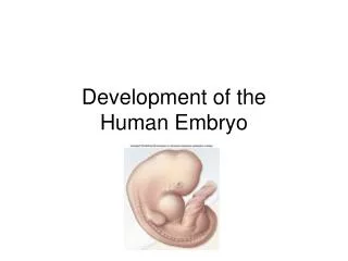
The Human Embryo
The Human Embryo. Preimplantation Biological and Ethical Observations. Science. Preimplantation Embryo Zygote to blastocyst. Preimplantation. Preimplantation Embryo travels along the genital tract before implantation A Period of intense molecular dialogue with the maternal environment.

The Human Embryo
E N D
Presentation Transcript
The Human Embryo Preimplantation Biological and Ethical Observations
Science Preimplantation Embryo • Zygote to blastocyst
Preimplantation Preimplantation Embryo travels along the genital tract before implantation • A Period of intense molecular dialogue with the maternal environment
Human Fertilization • Spermatazoon and Oocyte: • Mature male and female gametes • Each contain half the complete chromosomal complement • In fertilization, they fuse and a new individual originates with a complete genetic complement of 46 chromosomes
Gametes • In male and female derive from precursors called primordial germ cells • Their maturation process and differentiation is called gametogenesis • Maturation of the germs cells involves halving of the chromosomes in a process of nuclear division called meiosis • Meosis commences at puberty in the male, but female gamete maturation commences during foetal life
Meiosis http://www.youtube.com/watch?v=D1_-mQS_FZ0
Process of Fertilization • Human spermatazoon is 50 microns (one millionth of a meter) • From testicles to fallopian tube=7 meters • 100,000 times their length • During the journey spermatazoon undergoes capacitation that sets off the process of activation of the spermatazoon that permits it to meet and make contact with the oocyte • Movement depends on physical and chemical stimuli • Will survive a few days
Fertilization Mature Oocytes are large, even up to 150 microns; it contains all the material needed to begin embryonic growth
Fertilization Acrosome reaction, penetration of Zona pellucida and binding
Acrosome reaction permits sperm to penetrate the layers surrounding the oocyte and fuse with the zona pellucida Fertilization http://www.uchsc.edu/ltc/Fertilization.html
Fusion of gametes Fusion of gametes is an irreversible process that indicates the beginning of a new organism. The first consequence of the fusion is a chemical change in the fertilized oocyte, a sudden increase in the concentration of Ca2+ ions, forming an ionic calcium wave and activating the metabolic activity of the new individual.
Cortical reaction A new individual has been created who demonstrates a genetic and molecular pattern belonging to that of the human species. The cortical reaction prevents entry of other spermatazoa, protecting the new individual.
Formation of Pronuclei A new individual has formed. The male pronucleus is functionally active now, as is the female. Gender is already determined at penetration of the ZP. Many genes of the new individual are already active before the meeting of the pronuclei and have begun to have a key role in development.
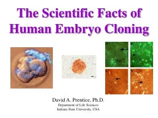
Pronuclei (in blue) coming towards each other during human fertilization. (A) Oocyte prior to fertilization; (B) penetration by the spermatozoon: the male pronucleus is on the left and the female pronucleus (which is completing its maturation) is on the upper right; (C) meeting and apposition of the pronuclei; preparation for the first cellular division: (D) prophase and (E) metaphase of the first mitotic division. The microtubules are marked in green and the DNA in blue (from SF. Gilbert, Developmental Biology, Sixth ed., Sinauer, Sunderland (MA) 2000, p.212, fig. 7-32).
First mitosis The paternal centrosome divides and organizes the mitotic spindle from the pronuclei; the duplicated male and female chromosomes move toward the equator of the spindle and are prepared for division.
Ordering of cell division Embryo at the stage of two cells marked in a differential way in order to follow the destiny of the descendant or daughter cells. (B) Blastocyst,embryonic pole –on top-, “abembryonic” pole –below- (from H. Pearson,Developmental Biology:Your destiny form day one, Nature 2002, 18, p.15).
ART and Genetic Diagnosis • Recent discoveries …highlight how potentially damaging operations on the early embryo can be for its subsequent development. • Certain techniques of assisted reproduction (for example, ICSI, Intra-Cytoplasmic Sperm Injection) …could destroy the delicate processes which allow the establishment of the body axes. • Even the genetic tests performed prior to implantation (Preimplantation Genetic Diagnosis), in which two cells are removed from the embryo at the stage of only eight cells, would appear to be another area of deep concern. Page 12 Human embryo in pre implantation stage.
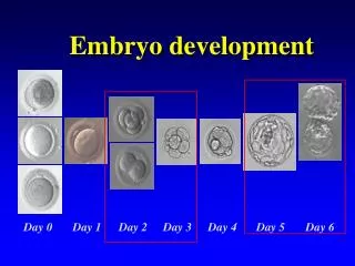
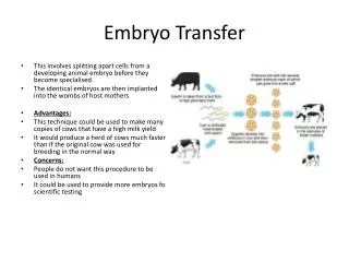
Development Prior to Implantation • For about 5 days cell division takes place called segmentation • The zygote is subdivided into smaller blastomeres • Forms a morula • 8-16 cell, compacts
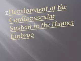
Developmental phases of the “preimplantation embryo”: the 2-cell stage(A); 4-cell stage (B); the morula, first at the 8-cell stage (C) and after compactionD, E); the blastocyst (F). (-Mouse embryo, in vitro development- by S. F. Gilbert,Developmental Biology, Sixth ed., Sinauer, Sunderland (MA) 2000, p. 356, fig.11-22).
Development prior to implantation • Much metabolic change responding to the energy needs • Apopstosis-programmed cellular death, removes abnormal and mutant cells • Growth factors, hormones, amino acids, carbs and proteins are produced by the embryo and regulate its development • Paf, a growth factor, stimulates metabolism and produces changes in the maternal environment
Maternal-Embryonic Dialogue • Oviduct is a reproductive organ which provides a series of molecules necessary for survival • The embryo produces hormones and other molecules necessary for interaction with the maternal environment • “Cross talk” prepares the embryo for implantation • On the 5th day the blastocyst comes out of the ZP, “hatching” and can now adhere to the uterine mucosa for implantation
Implantation Hatching allows embryonic individual to implant. This process is guided by cross talk between mother and developing individual.
Implantation Implantation commences with the apposition of microvilli of the uterine epithelium with those of the trophoblast which interlock with each other. Interactionbetween the trophoblast and the uterine epithelium requires cellular reorganisation, as mediated by a family of transmembrane receptors called Integrins, produced both by mother and embryo.
Implantation This relationship which begins at the moment of fertilization continues throughout pregnancy by biochemical, hormonal and immunological communication.
Prenatal and Preimplantation Diagnosis • Prenatal diagnosis-identifying pathologies before birth • Including chromosomal abnormalities • Mutations of one or more genes • Congenital diseases or malformations of an infective type • But the principal reason is genetic disease • There are some therapeutic possibilities but far fewer than the diseases that can be identified.
Invasive procedures • 1. Syncytiotrophoblast2. Trophoblastic lacunae3. Surface epithelium4. Fibrin coagulum5. Epiblast6. Aminotic cavity7. Hypoblast8. Cytotrophoblast9. Exocoelomic cavity Removal of foetal cells through the amniotic cavity
Invasive techniques • Analysis of chorionic villi: 10-14 weeks • Analysis of amniotic fluid: 15-16 weeks • Cordocentesis:18th week • Early amniocentesis: 11-14 weeks
Risk of foetal death increased • Villocentesis: 1-3% • Amniocentesis: 0.5-1% • A considerable percentage of premature births • Earlier techniques are associated with a greater risk of spontaneous abortion
Amniocentesis and abortion • Amniocentesis per year • US: 190,000 • France: 80,000 • Italy: 100,000 • In US, then 950 to 1,900 pregnancies with healthy babies end up with an abortion due to the technique. • Causes considerable stress and suffering to the foetus by changing the composition of the fluid
Noninvasive prenatal diagnosis • Searching for embryonic DNA in maternal blood-but limited by amount and the possibility of contamination with maternal cells • Ultrasound
Preimplantation diagnosis • Increase efficiency of IVF by selection • Preimplantation Genetic diagnosis • Removes cell after about 3 days • Chromosomal screening • High rate of diagnostic error 5-10% • Invasive • Cost $1500-3000 • The goal of the test? • Abortion • Sex selection • Eugenic