Download
1 / 32
370 likes | 587 Views
Cell Cycle Control Abnormalities result in cancer or developmental disorders. Cell Growth and Oncogenesis. Mechanisms Reduction in differentiation and/or PCD – no change in rate of proliferation Increased rate of proliferation without inhibition of differentiation/PCD. Cancer cells.

E N D
Cell Cycle Control Abnormalities result in cancer or developmental disorders
Cell Growth and Oncogenesis • Mechanisms • Reduction in differentiation and/or PCD – no change in rate of proliferation • Increased rate of proliferation without inhibition of differentiation/PCD Cancer cells Proliferation c40 divisions Cell number Normal cells - PCD Senescent DNA errors Time 100 1012 cells death 10 Cancer diameter (mm) 109 cells lump 1 0.1 108 cells X-ray visible 10 20 30 40 Divisions
Cancer and the Cell Cycle • Basic function to replicate exactly chromosomal DNA • DNA duplication occurs in S (synthesis) phase • Cell division in M phase • Gap phases G1 and G2
Phases of the Cell Cycle • G1 phase between M phase and S-phase • G2 phase S-phase and mitosis • G1 G2 S called the interphase • Monitoring and deletion function
Flow Cytometry of DNA content • DNA content in a cell population identifies cells with 2x content as dividing cells • Tumours are frequently aneuploid ie irregular cellular DNA content
S-phase identification • Thymidine incorporation • BrdU incorporation • Can be measured easily in whole animals and in tissue sections
The concept of the Check point • Multiple errors can occur in this process • Errors must be controlled • Elaborate machinery to control for errors • Genetic errors in error controlling machinery may be crucial
Cyclins • Originally identified in yeast • Tend to be evolutionarily conserved • Regulate progression through phases of the cell cycle • Act as regulatory units for cyclin dependent kinases CDKs • Cyclin –CDK complex regulated by phosphorylation
G0 G1 S G2 M P Rb P P Unphosphorylated Rb forms complex with E2F Rb E2F1 No transcription Phosphorylation of Rb (cycD-CDK4) promotes dissociation of E2F1 and Rb E2F1 Transcription Control of the Cell Cycle B A Interactions between cyclins and cyclin-dependent kinases regulate cycle D Cyclin Levels E cycB cycB cyc cycA cdc25 cdc2 CDK CDK2 cdc2 cycD cycE p27 Y pY CDK4 CDK2 p27 may regulate exit from G0 Phosphatase cdc25 dephosphorylates cdc2 allowing entry into M phase
P53 discovery SV40 T is a viral protein that transforms human cells
P53- “guardian of the genome” • P53 activated following genotoxic insult • Induces transcription of p21 • P21 locks CDK in the off position
P53 and cancer • Mutations cause Li Frameini syndrome • Multiple cancers in different organs • Somatic mutations often occur in cancer • Somatic mutations affect cell growth • Somatic mutations often signal progression to malignant phase
The p53 Signaling Pathway SIGMA-ALDRICH
Drugs may have phase-specific effects • Cytotoxic effects are frequently phase specific • Multiple phases may be targeted • Single stage may be targetted by multiple drugs
G1 and S Phases of the Cell Cycle SIGMA-ALDRICH CDK4
Why is this important? Colon Cancer CDK4 CDK4 modelling and drug design
G2 and M Phases of the Cell Cycle SIGMA-ALDRICH
Regulatory Cascade of Cyclin Gene Expression SIGMA-ALDRICH
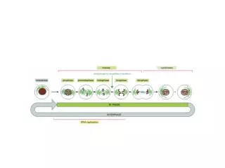
Structure of the cell cycle The cell cycle consists of four phases. Gap 1 (G1) is the interval between mitosis and DNA replication that is characterized by cell growth. If the conditions that signal transition to replicative phase are not present, the cell exits the cell cycle and enters G0, a nonproliferative phase during which growth, differentiation and apoptosis occur. DNA replication occurs during the synthesis (S) phase, which is followed by a second gap phase (G2) during which growth and preparation for cell division occurs. Mitosis occur in M phase.
G1 and S Phases of the Cell Cycle In proliferating cells, the cell cycle consists of four phases. Gap 1 (G1) is the interval between mitosis and DNA replication that is characterized by cell growth. The transition that occurs at the restriction point (R) in G1 commits the cell to the proliferative cycle. If the conditions that signal this transition are not present, the cell exits the cell cycle and enters G0, a nonproliferative phase during which growth, differentiation and apoptosis occur. Replication of DNA occurs during the synthesis (S) phase, which is followed by a second gap phase (G2) during which growth and preparation for cell division occurs. Mitosis and the production of two daughter cells occur in M phase. Passage through the four phases of the cell cycle is regulated by a family of cyclins that act as regulatory subunits for cyclin-dependent kinases (cdks). The activity of the various cyclin/cdk complexes that regulate the progression through G1 -S-G2 phases of the cell cycle is controlled by the synthesis of the appropriate cyclins during a specific phase of the cell cycle. The cyclin/cdk complex is then activated by the sequential phosphorylation and dephosphorylation of the key residues of the complex, located principally on the cdk subunits. The cyclin cdk complex of early G1 is either cdk2, cdk4, or cdk6 bound to a cyclin D isoform. There are several proteins that can inhibit the cell cycle in G1. If DNA damage has occurred, p53 accumulates in the cell and induces the p21-mediated inhibition of cyclin D/cdk. Mdm2, by facilitating the nuclear export/inactivation of p53, becomes part of an inhibitory feedback loop that inactivates p21-mediated G1 arrest. Similarly, activation of TGF- receptors induces the inhibition of cyclin D/cdk by p15, while cyclic-AMP inhibits the cyclin D/cdk complex via p27. If the cyclin D/cdk complex is inhibited, retinoblastoma protein (Rb) is in a state of low phosphorylation and is tightly bound to the transcription factor E2F, inhibiting its activity. Passage through the restriction point and transition to S phase is triggered by the activation of the cyclin D/cdk complex, which phosphorylates Rb. Phoshporylated Rb dissociates from E2F, which is then free to initiate DNA replication. Cyclin E/cdk2 accumulates during late G phase and triggers the passage into S phase. The entire genome is replicated during S phase. The synthesis and accumulation of cyclin B/cdc2 also begins during S phase, but the complex is phosphorylated at Thr14 -Tyr15 and is inactive. Cyclin A/cdk2 accumulates during S phase and its activation triggers the transition to G2, a phase characterized by the accumulation of cyclin B/cdc2, the inhibition of DNA replication, cell growth and new protein synthesis.
G2 and M Phases of the Cell Cycle The transition from G2 phase to mitosis is triggered by the Cdc25-mediated activation (dephosphorylation) of the cyclin B/cdc2 complex (MPF). The activation of cyclin B/cdc2 that is necessary for G/M progression is currently the most well-characterized step in the cell cycle. CyclinB/cdc2 is activated by phosphorylation of Thr160 and the dephosphorylation of Thr14 -Tyr15. Thr160 is phosphorylated by cyclin activating kinase (CAK), following the activation of CAK by a cyclin activating kinase activating kinase (CAKAK). However, the complex is kept in an inactive state due to the phosphorylation of Thr15, which is catalyzed by the Wee1 kinase. Cyclin B/cdc2 activation is triggered when Cdc25, a phospatase, dephosphorylates Thr15. In turn, the activity of Cdc25 is regulated by both activating and inhibitory phosphorylations. Phosphorylation of Ser by Chk1 (a check point activated kinase that participates in the G2-arrest of cells with damaged DNA) leads to the inactivation of Cdc25, while phosphorylation by an M-phase activated kinase creates a positive feedback loop leading to the rapid activation of the cyclin B/cdc2 complex. MPF catalyzes the phosphorylation of lamins and histone 1, and is involved in the regulation of events preceding cell division, such as spindle formation, chromatin condensation, and fragmentation of the nuclear envelope and of organelles such as the Golgi and endoplasmic reticulum. The metaphase to anaphase transition is triggered by inactivation of MPF and the degradation of cyclin B. This induces the separation of chromatids and their movement to the poles of the mitotic spindle, after which the mitotic apparatus disappears, the nuclear membranes reform and the nucleoli reappear. During cytokinesis, the cytoplasm divides and the resulting daughter cells enter G1. References: Smits, V.A., and Medema, R.H., Checking out the G2/M transition. Biochim.Biophys.Acta., 1519, 1-12 (2001). Taylor, W.R., and Stark, G.R., Regulation of the G2/M transition by p53. Oncogene, 20, 1803-1815 (2001). Bulavin, D.V., et al., p38 and Chk1 kinases: different conductors for the G2/M checkpoint symphony. Curr. Opin. Genet. Dev., 12, 92-97 (2002).
The p53 Signaling Pathway The tumor-suppressor protein p53 exhibits sequence-specific DNA-binding, directly interacts with various cellular and viral proteins, and induces cell cycle arrest in response to DNA damage. In response to signals generated by a variety of genotoxic stresses, e.g, UV irradiation or DNA damage, p53 is expressed and undergoes post-translational modification that results in its accumulation in the nucleus. The p53-dependent pathways help to maintain genomic stability by eliminating damaged cells, either by arresting them permanently or through apoptosis. For example, -irradiation activates p53 to turn on the transcription of p21CIP1, that, in turn, binds to and inhibits cyclin-dependent kinases, causing hypophosphorylation of retinoblastoma (Rb), thus preventing the release of E2F and blocking the G1-S transition. Some of the cellular effects of p53 can be blocked by the deregulated expression of c-Myc, Bcl-2, or E2F. p53 activity is controlled through an autoregulatory loop involving Mdm2. The binding of Mdm2 to p53 targets p53 for degradation and inhibits p53-induced cell-cycle arrest and apoptosis. References Gu, J., et al., Identification of a sequence element from p53 that signals for Mdm2-targeted degradation. Mol. Cell Biol., 20, 1243-1253 (2000). Jimenez, G.S., et al., p53 regulation by post-translational modification and nuclear retention in response to diverse stresses. Oncogene, 18, 7656-7665 (1999). King, K.L., and Cidlowski, J.A., Cell cycle regulation and apoptosis. Annu. Rev. Physiol., 60, 601-617 (1998).
Regulatory Cascade of Cyclin Gene Expression When cells traverse the G0 to G1 phase to the S-phase transition, a series of cyclin-dependent kinases is activated. The addition of serum growth factors to quiescent cells promotes transcription of the cyclin D1 gene. Cyclin D1 then associates with pre-existing cdk4 to form an active complex. The kinase activity associated with this complex can phosphorylate specific sites on the retinoblastoma protein (pRb), leading to inactivation of pRb and the activation cyclin E transcription by E2F. Activation of the cyclin E gene can be blocked by the cdk inhibitor p16. Cyclin E associates with existing cdk2 and this active complex regulates the function of several sets of target proteins. First, cyclin E/cdk2 complexes associate with E2F/p107 complexes to activate expression of the cyclin A gene. Also, cyclin E/cdk2 complexes cooperate with cyclin D1 to amplify the phosphorylation of pRb. Cyclin A associates with cdk2 to form a kinase complex that phosphorylates downstream targets involved in the initiation of DNA replication. References Ohtani, K., Implication of transcription factor E2F in regulation of DNA replication. Front. Biosci., 4, D793-D804 (1999). Lania, L., et al., Transcriptional control by cell-cycle regulators: a review. J. Cell. Physiol., 179, 134-141 (1999). Hatakeyama, M., et al., The role of RB in cell cycle control. Prog. Cell Cycle Res., 1, 9-19 (1995).