Download
1 / 13
130 likes | 403 Views
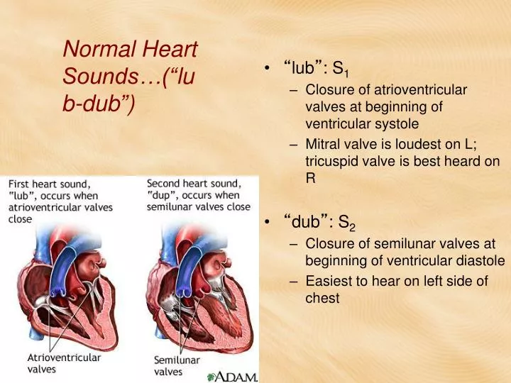
Normal Heart Sounds…( “ lub-dub ” ). “ lub ” : S 1 Closure of atrioventricular valves at beginning of ventricular systole Mitral valve is loudest on L; tricuspid valve is best heard on R “ dub ” : S 2 Closure of semilunar valves at beginning of ventricular diastole

E N D
Normal Heart Sounds…(“lub-dub”) • “lub”: S1 • Closure of atrioventricular valves at beginning of ventricular systole • Mitral valve is loudest on L; tricuspid valve is best heard on R • “dub”: S2 • Closure of semilunar valves at beginning of ventricular diastole • Easiest to hear on left side of chest
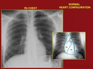
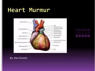
Valve Locations P = Pulmonic valve A = Aortic valve M = Mitral valve T = Tricuspid valve
Normal Heart Sounds (“lub-dub”) • Large animals - additional heart sounds • S3 - Rapid ventricular filling • S4 - Contraction of atria • not usually heard in small animals
Vascular Anatomy and Physiology • Arteries—carry blood away from heart • Veins—carry blood toward heart Blood in the systemic circulation is under higher pressure than blood in the pulmonary or coronary circulation. More pressure is needed to carry blood throughout the body. The systemic circulation encounters more resistance to flow. With the exception of the pulmonary arteries and veins and the umbilical arteries and veins, arteries carry oxygenated blood and veins carry deoxygenated blood.
Vascular Anatomy • Aorta- Largest artery in body, largest diameter and thickest vessel walls. • Arterial walls layers are similar to the layers of the heart. • Tough outer fibrous layer • Middle layer of smooth muscle and elastic connective tissue. • Smooth inner lining called endothelium In aorta and pulmonary arteries, the middle layer contains more elastic fibers- allows them to stretch slightly as they receive the high-pressure blood from the ventricles.
Vascular Anatomy Continued • Smaller arteries continue to split into smaller and smaller vessels and then arterioles • Blood flows through arterioles into tiny, thin-walled capillaries • Capillaries have no muscle layer in their wall, which are made up of only one cell layer (simple squamous epithelium). It is easy for the blood to exchange oxygen and nutrients for carbon dioxide and vice versa
Vascular Anatomy Continued • Blood travels back to heart through small venules which merge to form veins • Venous blood is under lower pressure than arterial blood • Veins have thinner walls than arteries • Valves in veins ensure that blood travels only toward heart • Veins usually are located next to arteries.
Vascular Anatomy Continued • Smooth muscle in walls of most blood vessels • Constriction and relaxation allows vascular system to direct blood to different regions of the body under different circumstances
Vascular Anatomy • Right and left subclavian arteries branch off of the aorta and travel to forelimbs. • Cartoid arteries branch off one or both subclavian arteries and supply blood to the head. • Main trunk of aorta arches dorsally and then travels caudally just below the spine. • Numerous branches emerge in the thoracic and lumbar areas • At hind limbs, aorta branches into right and left iliac arteries which supply hindlimbs. • Small coccygeal artery emerges to supply blood to tail.
Vascular Anatomy Continued • Jugular veins drain blood from the head. • Veins in the foreleg merge into larger and larger vessels to form right and left brachiocephalic veins • These carry blood to the cranial vena cava then back into the right atrium. • Veins in the hind limbs merge into right and left iliac veins • These carry blood to the caudal vena cava. • Caudal vena cava travels to the right atrium
Venipuncture • Jugular Vein: Ventral aspect of each side of the neck. • Close to the carotid arteries • Care must be taken to avoid accidental injection into the carotid artery • Cephalic vein: craniomedialaspect of forelimb. • Femoral Vein: medial aspect of hind limb. • Saphenous: lateral aspect of hind limb.
Venipuncture • Caudal epigastric vein: also called Milk vein. • found superficially on the ventral aspect of each side of the abdomen in lactating cows • not generally used for venipunture since it is thin-walled and prone to hematoma formation. • Coccygeal vein: also called Tail vein. • runs along ventral midline of the tail • Used for venipuncture in rodents and ruminants