The Skeletal System
280 likes | 292 Views
This text provides an overview of the skeletal system, including its structure, functions, and common disorders. Learn about the different types of bones, bone development and remodeling, and the role of joints in movement. Discover how the skeletal system supports the body, protects organs, and facilitates motion. Explore the various bones of the human body, including the skull, vertebral column, and appendages. Gain insight into joint classifications and the clinical forms of arthritis.

The Skeletal System
E N D
Presentation Transcript
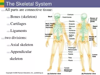
The Skeletal System • All parts are connective tissue: • Bones (skeleton) • Cartilages • Ligaments • two divisions: • Axial skeleton • Appendicular skeleton
Functions of Skeleton and Bones • Support of the body • Protection of soft organs • Movement due to attached skeletal muscles • Storage of minerals and fats • Blood cell formation
Bones of the Human Body • 206 bones • Two basic types • Compact bone • Dense • Calcium crystals • Spongy bone • Trabeculae - “little beams” • Many open spaces Figure 5.2b
Gross Anatomy of a Long Bone Epiphysis • Ends of the bone - spongy bone • Red bone marrow Diaphysis • Shaft - compact bone • Medullary cavity Periosteum Figure 5.2a
Structure of Bone Osteon - cylinder of bone, has central canal with blood vessels Osteocytes within spaces (lacunae), connected by small canals (canaliculi) Figure 5.1
Cartilage and Ligaments • Ligaments - dense fibrous connective tissue • Collagen fibers, fibroblasts • Cartilage - collagen and elastin fibers in base of water. Smooth and flexible substance • used to reduce friction, provide support under pressure at joints
Changes in the Human Skeleton • Embryos - hyaline cartilage • Cartilage replaced by bone = ossification • done by osteoblasts - bone building cells • Secrete osteoid (proteins, fibers, enzymes) • Osteoid replaced by calcium phosphate salts • Cartilage remains in nose, ribs, joints, and epiphyseal plates, ends of bones
Bone Development (cont.) Figure 5.2
Types of Bone Cells • Chondrocytes • Cartilage-forming cells • Osteocytes • Mature bone cells • Osteoblasts • Bone-forming cells • Osteoclasts • Bone-destroying cells • Break down bone matrix for remodeling and release of calcium
Mature Bone Remodeling and Repair • Changes in shape, size, strength • Dependent on diet, exercise, age • Bone cells regulated by hormones • Parathyroid hormone (PTH): removes calcium from bone • Calcitonin: adds calcium to bone
Stages in the Healing of a Bone Fracture Fibrocartilage callus is replaced by a bony callus • Bony callus is remodeled to form a permanent patch • Hematoma (blood-filled swelling) is formed • Break is splinted by fibrocartilage to form a callus Figure 5.5
The Axial Skeleton • Forms the longitudinal part of the body • Divided into 3 parts • Skull • Vertebral column • Bony thorax
The Skull • Two sets of bones • Cranium • Facial bones • Bones are joined by sutures • Only the mandible is attached by a freely movable joint
The Hyoid Bone • only bone that does not articulate with another bone • Serves as moveable base for the tongue Figure 5.12
Paranasal Sinuses • Functions of paranasal sinuses • Lighten the skull • Give resonance and amplification to voice Figure 5.10
The Fetal Skull • Fontanelles – fibrous membranes connecting the cranial bones • Allow the brain to grow • Convert to bone within 24 months after birth Figure 5.13
The Vertebral Column • 5 regions, 33 bones • Spine has a normal curvature • Vertebrae separated by intervertebral discs Figure 5.7
The Bony Thorax • Made-up of two parts • Sternum • Ribs (12 pr) • Forms a cage to protect major organs Figure 5.19a
The Appendicular Skeleton • Limbs (appendages) • Pectoral girdle • Pelvic girdle
Bones of the Pectoral Girdle • Clavicle – collarbone • Scapula – shoulder blade Figure 5.20a–b
Bones of the Upper Limb • The arm - Humerus • The forearm - Ulna and Radius Hand - Carpals Metacarpals Phalanges Figure 5.21a–b
The Pelvis Figure 5.23a
Gender Differences of the Pelvis Figure 5.23c
Bones of the Lower Limbs Femur – thigh bone Tibia - bears weight Fibula - helps rotation with ankle Tarsals - ankle Metatarsals - foot Phalanges - toes Figure 5.24a–b
Joints (Articulations) • Classified by degree of movement • Fibrous joint: immovable; e.g., fontanels • Cartilagenous joint: slightly movable, cartilage connection; e.g., backbone • Synovial joint: freely movable
Synovial Joints - movable; all limbs • Joint capsule: synovial membrane + hyaline cartilage • Synovial membrane secretes synovial fluid as lubricant • Hyaline cartilage cushions
Disorders, Inflammatory Conditions Associated with Joints • Sprains: stretched or torn ligaments • Bursitis and tendinitis: inflammations
Clinical Forms of Arthritis • Osteoarthritis • Cartilage wears out, pain, inflammation • normal aging processes • Rheumatoid arthritis • autoimmune disease –attacks the joints • inflammation of synovial membranes • Cartilage lost, bones fused, leads to deformities


