Download
1 / 46
500 likes | 831 Views
Chapter 15 Cardiovascular System. heart blood vessels. Average Size of Heart cm long cm wide. 15-2. Location of Heart. posterior to sternum medial to lungs anterior to vertebral column. 15-3. Coverings of Heart. 15-4. Wall of Heart. Three layers

E N D
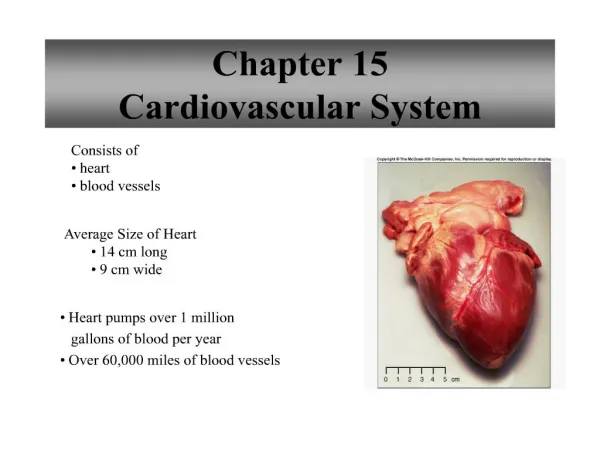
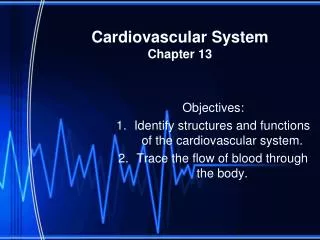
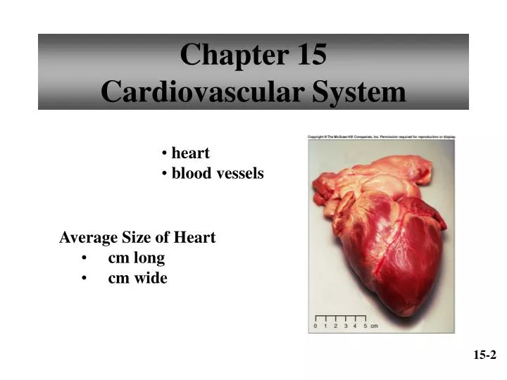
Chapter 15Cardiovascular System • heart • blood vessels • Average Size of Heart • cm long • cm wide 15-2
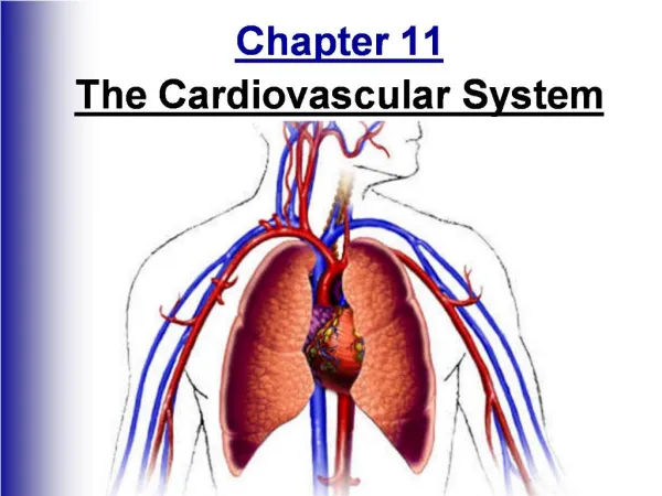
Location of Heart • posterior to sternum • medial to lungs • anterior to vertebral column 15-3
Coverings of Heart 15-4
Wall of Heart • Three layers • forms protective inner lining • cardiac muscle • contracts to pump blood • serous membrane • protective covering • contains capillaries and nerve fibers 15-5
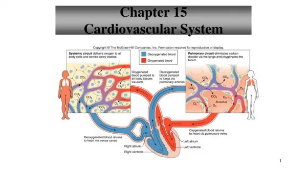
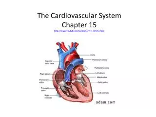
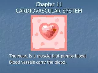
Heart Chambers • Right Atrium • inferior vena cava • superior vena cava • Left Atrium • Right Ventricle • Left Ventricle 15-6
Heart Valves • Tricuspid Valve • right A-V valve • Bicuspid Valve • left A-V valve • Aortic Valve • semilunar valve • Pulmonary Valve • semilunar valve 15-7
Heart Valves Tricuspid Valve Pulmonary and Aortic Valve 15-9
Skeleton of Heart • fibrous rings to which the heart valves are attached 15-10
Path of Blood Through the Heart Pumps your blood 15-11
Blood Supply to Heart 15-14
Cardiac Cycle • Atrial Systole/Ventricular Diastole • remaining 30% of blood pushed into ventricles • ventricles relaxed • Ventricular Systole/Atrial diastole • chordae tendinae prevent cusps of valves from bulging too far into atria • blood flows into atria • blood flows into pulmonary trunk and aorta Cardiac Cycle.exe 15-17
Heart Sounds • Lubb • first heart sound • occurs during ventricular contraction Normal Beat.mov • Dupp • second heart sound • occurs during ventricular contraction Murmur – Murmer.mov 15-18
Heart Sounds 15-19
Cardiac Conduction System 15-22 Conducting System of the Heart.exe
Electrocardiogram • recording of electrical changes that occur in the myocardium • used to assess heart’s ability to conduct impulses P wave – atrial depolarizatoin QRS wave – ventricular depolarization T wave – ventricular repolarization 15-24
Electrocardiogram 15-25
Electrocardiogram A prolonged QRS complex may result from damage to the A-V bundle fibers 15-26
Regulation of Cardiac Cycle • physical exercise • body temperature • parasympathetic impulses decrease heart action • sympathetic impulses increase heart action • cardiac center regulates autonomic impulses to the heart 15-28
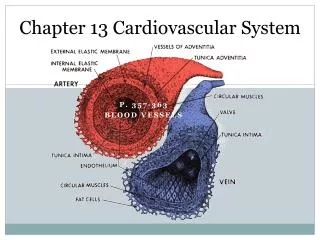
Blood Vessels • arteries • arterioles • capillaries • venules • veins 15-30
Arteries and Arterioles • Arterioles • thinner wall than artery • helps control blood flow into a capillary • Artery • thick strong wall Heart Attach.mov 15-31
Walls of Artery and Vein 15-32
Arteriole • smallest arterioles only have a few smooth muscle fibers • capillaries lack muscle fibers 15-33
Capillaries • extensions of inner lining of arterioles 15-35
Exchange in the Capillaries • water and other substances leave capillaries because of net outward pressure at the capillaries’s arteriolar ends • water enters capillaries’s venular ends because of a net inward pressure 15-38
Venules and Veins • Venule • thinner wall than arteriole • less smooth muscle and elastic tissue than arteriole • Vein • three layers to wall but middle layer is poorly developed • serves as blood reservoir 15-39
Venous Valves 15-40
Blood Volumes in Vessels 15-41
Arterial Blood Pressure Blood Pressure – • Arterial Blood Pressure • rises when ventricles contract • falls when ventricles relax • systolic pressure – • diastolic pressure – 15-42
Pulse • alternate expanding and recoiling of the arterial wall that can be felt 15-43
Pulmonary Circuit • consists of vessels that carry blood from the heart to the lungs and back to the heart 15-50
Blood Flow Through Alveoli • cells of alveolar wall are tightly joined together • the high osmotic pressure of the interstitial fluid draws water out of them 15-51
Systemic Circuit • includes the aorta and its branches • includes the system of veins that return blood to the right atrium 15-52
Life-Span Changes • deposition of cholesterol in blood vessels • heart enlarges • cardiac muscle cells die • fibrous connective tissue of heart increases • adipose tissue of heart increases • blood pressure increases • resting heart rate decreases 15-70
Clinical Application Arrhythmias • Ventricular fibrillation • rapid, uncoordinated depolarization of ventricles • Tachycardia • rapid heartbeat • Atrial flutter • rapid rate of atrial depolarization 15-71
Angiogram • Angioplasty & Stenting • Atherectomy • Bypass Surgery • Coronary Artery Disease • Plaque Rupture