Download
1 / 23
250 likes | 357 Views
Explore the complexities of pulmonary respiration, from the anatomy of the respiratory system to the exchange of gases in the lungs. Learn about the functions, types, and clinical applications of respiration in this comprehensive guide.

E N D
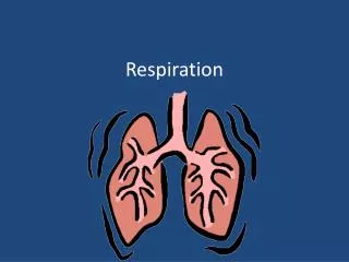
Respiration And the Pulmonary System
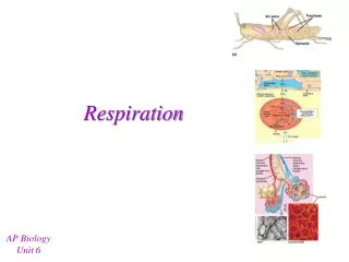
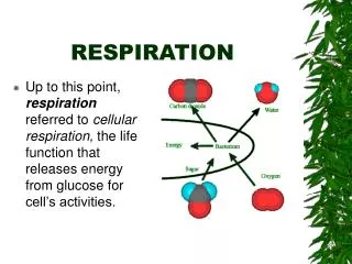
Pulmonary respiration (ventilation) – Breathing Inspiration Expiration External respiration – between lungs and blood Internal respiration – Between blood and cells Cellular respiration Glucose + Oxygen Carbon Dioxide and water and energy. Types of Respiration
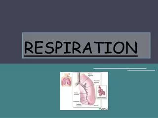
By location Upper respiratory system Nose Pharynx and associated structure Lower respiratory system Larynx Bronchial tree Lungs By function Conducting portion Nasal passageways Pharynx Larynx Respiratory portion Bronchial tree – Bronchi terminal bronchiole Respiratory bronchioles Alveolar ducts Alveoli Organization of Respiratory Organs
Components External Nasal bones Alar cartilage External nares – Nostils Nasal septum Internal Choanae Internal nares Mucous membrane Paranasal sinuses Frontal Sphenoidal Ethmoidal Maxillary Functions Incoming air Warmed Moistened Filtered Olfactory stimuli received Sound Resonate Modification Nose
Extent Internal nares Cricoid cartilage (larynx) Regions Nasopharynx Openings Internal nares Auditory (Eustachian) tubes Pharyngeal tonsil (adenoid) Oropharynx Opening – Fauces Tonsils Palatine Lingual Common Path Air Drink Food Laryngopharynx (hypopharynx) – Connected inferiorly Esophagus Larynx Pharynx (throat)
Joins pharynx to trachea Cartilages 3 unpaired Epiglottis Protects airway Covers glottis Thyroid – Adam’s apple Cricoid – Tracheostomy landmark 3 paired Arytenoid Corniculate Cuneiform Voice production Laryngeal mucous membranes Ventricular folds (false vocal chords)– Superior Vocal folds (true vocal chords) – Inferior Bring folds together Hold breath against pressure Vibrate in response to pressure Larynx (Part 1)
Control Loudness – Air pressure Pitch – vocal fold tension Resonance Upper respiratory tract Paranasal sinuses Modifications – Muscles Pharynx Face Tongue Cheeks Larynx (Part 2)
Windpipe Leads from larynx into bronchial tree Sternal angle T5 Carina – Cough reflex C-shaped cartilage Holds trachea open Allows esophageal expansion Clinical applications Tracheostomy Intubation Trachea
Begin at sternal angle (T5) Diameter decreases as branching increases Amount of cartilage decreases as diameter decreases Amount of smooth muscle increases as diameter increases Primary (serve a lung) Right Wider diameter Shorter More vertical Left Smaller diameter Longer More horizontal Secondary (serve a lobe) 3 on right 2 on left Tertiary – Segmental or lobular Bronchi (Part 1)
Tertiary – Segmental or lobular Bronchioles Small branches of bronchial area Terminal – Extend into alveolar clusters Respiratory – Extend directly into alveoli ANS effects Sympathetic --Bronchodilate Parasympathetic -- Bronchoconstrict Bronchi (Part 2)
Enclosed by pleurae Parietal Visceral Pleural cavity Gross anatomy Base – fits over diagragm Apex – extends into root of neck Costal surface – Lies against ribs Mediastinal surface Faces heart Hilus (hilum) – Entrance/Exit Blood vessels Bronchi Nerves Right lung 3 lobes Superior Middle Inferior 2 fissures Lungs (Part 1)
Oblique Horizontal Left lung 2 lobes Superior Inferior 1 fissure – Oblique Cardiac notch Lungs (Part 2)
1 respiration = 1 inspiration + 1 expiration Exchange of gases between atmosphere and lungs Normal inspiration (inhalation) Increase thoracic cavity volume – Contract Diaphragm External intercostals Reduction in intrapleural pressure Air rushes into lungs Forced inspiration Body needs more air exchange Need more change in thoracic cavity volume Use additional muscles to raise thoracic cage Sterrocleidomastoid Scalenes Pectoralis minor Pulmonary Ventilation - Respiration (Part 1)
Normal expiration (exhalation) Decrease thoracic cavity volume Diaphragm relaxes Intrapleural pressure increases Air pushed out of lungs Forced expiration Body needs more air exchange Active process using Abdominal muscles Internal intercostals Factors affecting ease of respiration Compliance Elasticity Surface tension – Surfactant Airway resistance Modified respirations Cough Sneeze Sigh Yawn Pulmonary Ventilation - Respiration (Part 2)
Laugh Hiccuping Related terminology Hyperventilation Hypoventilation Eupnea Dyspnea Apnea Shortness of Breath (SOB) Atelectasis Pulmonary Ventilation - Respiration (Part 3)
Lung – Lobe – Segment – Lobule – Alveoli Alveolus Epithelial “bubble” Type I cells – lining Type II cells – surfactant Alveolar macrophages Monocytes Fibroblasts Alveolar capillary membrane Respiratory membrane Components Alveolar wall Epithelial basement membrane Capillary basement membrane Capillary endothelial Thickness – 0.5 microns Allows fast exchange of respiratory gases Total surface area – 70 square meters (750 square feet) Lung Histology
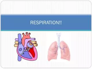
Bronchial Arteries Bring blood to supply lung cells Veins Drain blood from lung cells Drain into azygous system Pulmonary Arteries Carry oxygen poor blood fromR. Ventricle for perfusion Veins Carry oxygen rich blood back to L. ventricle for systemic circulation Lung Blood Supply
Oxygen Very little dissolved in plasma Most bound to hemoglobin (Hb) 1 O2/heme 4 hemes/Hb Hb+O2 HbO2 Carbon dioxide Small amount dissolves in plasma – More soluble than oxygen Carbaminohemoglobin – Hb + CO2 HbCO2 As bicarbonate ions CO2+H2OH2CO3 H2CO3H+HCO3 Respiratory Gases in the Blood
1 respiration = 1 inspiration + 1 expiration Should be About 12 per minute About 6 L per minute Measure with spirometer Pulmonary volumes (specific conditions) Tidal volume (TV) Minute respiratory volume (MVR) – TV x respiration rate Inspiratory reserve volume (IRV) Expiratory reserve volume (ERV) Residual volume (RV) Minimal volume (MV) Pulmonary capacities (combined conditions) Inspiratory capacity – TV +IRV Function residual capacity – RV+ERV Vital capacity – IRV+TV +ERV Total capacity – TV+IRV+ERV+RV+MV Pulmonary Function Measurements
Respiratory centers Medullary rhythmicity Areas Inspiration Expiratory Sets basic rhythm 2 sec inspiration 3 sec expiration Communicate with diaphragm Phrenic n. Intercostal n. Pons Helps switch between inspiration/expiration Areas Pneumotaxic Limits inspiration Overrides apneuistic area Apneuistic Limits expiration Stimulates inspiration Works when pneumotaxis area is inactive Control of Respiration (Part 1)
Influencing factors Vagus n. Bronchial stretch receptors – Inflation reflex Anal sphincter receptors Chemical stimuli Medulla oblongata – Central chemoceptors – H ions Peripheral chemoceptors Where Aortic body Carotid body What H ions CO2 O2 Proprioceptors Increased body temperature Pain Acute Chronic Upper respiratory irritation Emotional stimuli Cortical influences Control of Respiration (Part 2)