Download
1 / 42
430 likes | 715 Views
Protein Folding Purification and Myoglobin Lecture 11 (29 September 2009). Protein Folding. Protein folding problem. Levinthal paradox 100aa protein three conformations => 3 100 possible orientations

E N D
Protein Folding Purification and MyoglobinLecture 11(29 September 2009)
Protein Folding Protein folding problem • Levinthal paradox • 100aa protein three conformations • => 3100 possible orientations • => random search for native structure would take longer than the age of the universe • Prediction of three dimensional structure from its amino acid sequence • Translate “Linear” DNA Sequence data to spatial information
Sidechain locations in proteins • Non-polar sidechains (Val, Leu, Ile, Met, and Phe) occur mostly in the interior of a protein keeping them out of the water (hydro-phobic effect) • Charged polar residues (Arg, His, Lys, Asp, and Glu) are normally located on the surface of the protein in contact with water. • Uncharged polar residues (Ser, Thr, Asn, Gln, and Tyr) are usually on the protein surface but also occur in the interior of the protein.
Protein Stability Forces that stabilize protein structure: 1, 2, 3 1. The Hydrophobic Effect
2. Electrostatic Interactions Ion pair (salt bridge) of myoglobin 3. Chemical Cross-links Zinc finger: Nucleic acid-binding proteins
Protein Folding Pathways Proteins can be unfolded/denatured. Denatured proteins can be refolded, sometimes requiring helper proteins, and this refolding takes place via preferred pathways. Common thought is that secondary structures form first, eventually collapsing due to the formation of hydrophobic cores. Folding funnel Energy-entropy relationship for protein folding
GroEL GroES Molecular chaperons Molecular chaperones: (1) Hsp70 proteins function as monomer (2) Chaperonins, large multisubunit proteins (3) Hsp90 proteins for the folding of proteins involved with signal transduction
Reaction cycle of the GroEL/ES cycle 1. GroEL ring binding 7 ATP and a substrate (improperly folded protein). Then it binds a GroES cap to become the cis ring. 2. The cis ring catalyzes the hydrolysis of its 7 ATP. 3. A 2nd substrate binds to the trans ring followed by 7 ATP. 4. The binding of substrate and ATP to the trabs ring conformationally induces the cis ring to release its bound GroES, 7 ADP, and the better folded substrate.The trans ring becomes the cis ring.
Diseases Caused by Protein Misfolding Alzheimer’s disease Transmissible spongiform encephalopathies (TSE) Amyloidoses Prion protein conformation
Once it has formed, an amyloid fibril is virtually indestructible (interchain H- bonds). It seems likely that protein folding pathways have evolved not only to allow polypeptides to assume stable native structures but also to avoid forming interchain H-bonds that would lead to fibril formation . The factors that trigger amyloid formation remain obscure, even when mutation (hereditary amyloidoses) or infection (TSEs) appear to be the cause. A model of an amyloid fibril
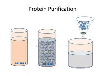
Protein Purification and Analysis General approach to purifying proteins Protein solubility Chromatography Electrophoresis Ultracentrifugation
Characteristic Charge Polarity Size Specificity Solubility Procedure 1. Ion exchange, 2. Electrophoresis, 3. Isoelectric focusing 1. Adsorption chromatography 2. Paper chromatography 3. Reverse phase chromatography 4. Hydrophobic interaction 1. Dialysis and ultrafiltration, 2. Gel electrophoresis, 3. Gel filtration, 4. Ultracentrifugation 1. Affinity chromatography 2. Immunopurification 1.Salt precipitation 2. Detergent solubilization Strategy of Purification Fractionation procedures or steps to isolate protein based on physical/chemical characteristics.
Protein Solubility • Since proteins contain a number of charged groups, its solubility depends on the concentration of dissolved ions • Salting in • At low ionic strength, increases in the concentration of dissolved ions leads to an increase in solubility by weakening the interaction between individual protein molecules. Interactions between protein molecules leads to aggregation (i.e. insolubility of proteins. • Salting out • As the ionic strength increases, they out compete the proteins for water molecules and the proteins become less soluble, aggregate, and fall out of solution.
a). At low ionic strength, all of the proteins are soluble b). As the ionic strength increases, the least soluble protein precipitates c). At even higher ionic strengths, further proteins precipitate. This process is continued until the desired protein is precipitated. This process not only allows you to obtain the desired protein, it removes many unwanted proteins in the process • Proteins are least soluble when they are neutral, so these salting out experiments are usually carried out at the pI of the protein (i.e. the isoelectric point where pH=pI, and the net charge on the protein is 0)
Salting out Use (NH4)2SO4 : it is a Very Soluble salt that does not harm proteins.
Solubility of b-lactoglobulin as a function of pH Solubility of carboxy-hemoglobin at its isoelectric point
Chromatography • Analytical methods used to separate molecules. Involves a mobile and a stationary phase. • Mobile phase is what the material to be separated is dissolved in. • Stationary phase is a porous solid matrix which the mobile phase surrounds. • Separation occurs because of the differing binding/ interactions each molecule has with both the mobile and stationary phase. • Interactions are different depending on the specific method.
Types of chromatography • Gas - liquid: Mobile phase is gaseous, stationary phase is liquid usually bound to a solid matrix. • Liquid - Liquid: Mobile phase is liquid, stationary phase is liquid usually bound to a solid matrix. • If separation is based on ionic interaction the method is called Ion Exchange Chromatography. • If separation is based on solubility differences between the phases the method is called Adsorption Chromatography. • If the separation is base on size of molecule the method is called Gel Filtration or Size Exclusion. • If the separation is base on ligand affinity the method is called Affinity Chromatography.
Ion ExchangeChromatography • A solid matrix with a positive charge, i.e., R+ can bind different anions with different affinities. • We can swap one counter ion for another • (R+A-) + B- (R+B-) + A- • R = Resin and exchanges Anions (-) • This is an anion exchange resin – the stationary phase is decorated with positively charged groups which bind anions in the mobile phase • There are also cation exchange resins. The type of an R group can determine the strength of interaction between the matrix, R and the counter ion. • If R is R- • (R-A+) + B+ (R-B+) + A+
Proteins have a net charge • The charge is positive below pI, • while the charge is negative above pI • The choice of exchange resin depends on the charge of the protein and the pH at which you want to do the purification. • Once the protein binds, all unbound proteins are washed off the column. Bound proteins are eluted by increasing the ionic strength, changing the counter ion or changing the pH altering the charge on the protein or the column.
The tan region is the ion exchange resin The mixture of proteins is the purple disc in a). The salt concentration is low at the beginning so the proteins with the lowest affinity for the column go through first (red protein) The salt concentration is then increased, washing off the proteins that interact more strongly with the ion exchange medium in the column Ion Exchange Chromatography • The most frequently used anion exchanger is: diethylaminoethyl (DEAE) • Matrix-CH2-CH2-NH(CH2CH3)2+ • The most frequently used cation exchanger is: carboxymethly • Matrix-CH2-COO-
Each gel bead consists of a gel matrix (wavy lines in the brown spheres) Small molecules (red dots) can fit into the internal spaces in the beads and get stuck Larger molecules (blue dots) cannot fit into the internal spaces in the beads and they come through the column faster Gel Filtration Chromatography
Gel filtration can be used to determine the molecular mass of proteins
Ligands (yellow in the figure to the left) are attached to the solid resin matrix The proteins in the eluant have ligand binding sites, however, only one of them will have the binding site for the ligand attached to the solid resin matrix The proteins that do not have the proper ligand binding site will flow through the column fastest The desired protein (i.e. the one with the proper ligand binding site) is then recovered from the column by washing with a solution with high ligand concentration, altered ionic strength, or altered pH Affinity Chromatography
Affinity Chromatography Based on molecular complementary between an enzyme and substrate. The substrate (R) is linked to a matrix with a spacer arm Only protein that binds R will stick to column. Put citrate on column citrate dehydrogenase will specifically bind. Add excess citrate and the enzyme will be released.
Electrophoresis is a method for separating proteins based on how they move in an electric field Image to the left is an electrophoretogram of serum, stained with amido black The sample starts at the top, an electric field is applied, and proteins migrate The molecules at the bottom are the lightest Molecules of similar charge and size move through the gel as a band The pH is typically 9 in these experiments so most proteins have a net negative charge and move toward the positive electrode (i.e. the one attached to the bottom of the gel) Gels are typically made of polyacrylamide and so the experiment is called polyacrylamide gel electrophoresis (PAGE) Electrophoresis
Sodium dodecyl sulfate (SDS) polyacrylamide gel electrophoresis (PAGE), SDS-PAGE is used to separate protein mixtures in a protein denaturing environment (SDS – soap) That is, the SDS causes proteins to denature and take on a rodlike shape and have similar charge to mass ratios Therefore, proteins are separated by molecular mass Again, the lighter proteins travel further In the figure, several (8) protein mixtures are run at the same time, some are controls and the others are samples Each sample is in a separate column, called a “lane” SDS-PAGE
A centrifuge is an instrument that rotates, generating centrifugal fields in excess of 600,000 times that of gravity This causes molecules in solution to undergo sedimentation at different rates, which are related to their masses The rate of sedimentation is measured in “s” which is the sedimentation velocity per unit of centrifugal force They are normally expressed in units of “S” (Svedbergs). One Svedberg is 10-13s Proteins: 1-50S Viruses: 40-1000S Organelles: tens of thousands of S Ultracentrifugation
Lysate - broken (lysed) cells- can be separated using • differential centrifugation • RPM - “spun down” • separates by density differences or by size (MW) of particles. • Cellular fractionation • can separate • mitochondria • microsomes • ribosomes • soluble proteins
Myoglobin and Hemoglobin • Because of its red color, the red blood pigment has been of interest since antiquity. • First protein to be crystallized - 1849. • First protein to have its mass accurately measured. • First protein to be studied by ultracentrifugation. • First protein to associated with a physiological condition. • First protein to show that a point mutation can cause problems. • First proteins to have X-ray structures determined. • Theories of cooperativity and control explain hemoglobin function
The Backbone structure of Myoglobin 33 Myoglobin: 44 x 44 x 25 Å single subunit 153 amino acid residues 121 residues are in an a helix. Helices are named A, B, C, …F. The heme pocket is surrounded by E and F but not B, C, G, also H is near the heme. Amino acids are identified by the helix and position in the helix or by the absolute numbering of the residue.
The Heme group Each subunit of hemoglobin or myoglobin contains a heme. - Binds one molecule of oxygen - Heterocyclic porphyrin derivative - Specifically protoporphyrin IX The heme prosthetic group in Mb ad Hb: protoporphyrin IX + Fe(II) The iron must be in the Fe(II)form or reduced form (ferrous oxidation) state.
Helix E Distal His Proximal His Helix F • Role of the Globin • Modulate oxygen binding affinity • Make reversible oxygen binding possible By introducing steric hindrance on one side of the heme plane interaction can be prevented and oxygen binding can occur. FeO OFe A heme dimer is formed which leads to the formation of Fe(III)
E7 F8
The visible absorption spectra for hemoglobin The red color arises from the differences between the energy levels of the d orbitals around the ferrous atom. Fe(II) = d6 electron configuration low spin state Binding of oxygen rearranges the electronic distribution and alters the d orbital energy. This causes a difference in the absorption spectra. Bluish for deoxy Hb Redish for Oxy Hb Measuring the absorption at 578 nm allows an easy method to determine the percent of O2 bound to Hb
Hemoglobin Spherical 64 x 55 x 50 Å two fold rotation of symmetry a and b subunits are similar and are placed on the vertices of a tetrahedron. There is no D helix in the a chain of hemoglobin. Extensive interactions between unlike subunits a2-b2 or a1-b1 interface has 35 residues while a1-b2 and a2-b1 have 19 residue contact. Oxygenation causes a considerable structural conformational change
Quaternary structure of deoxy- and oxyhemoglobin R-state T-state