Syncope: Guidelines & Risk Assessment 2017
230 likes | 250 Views
Explore the 2017 guideline on Syncope, the sudden loss of consciousness due to cerebral hypoperfusion, and differentiate it from other causes of Transient Loss of Consciousness (T-LOC). Learn about the various etiologies, risk assessment, and management strategies outlined in the ACC/AHA guideline. Dive into the evaluation process, high-risk factors, diagnostic work-ups, and imaging recommendations for patients experiencing syncope episodes.

Syncope: Guidelines & Risk Assessment 2017
E N D
Presentation Transcript
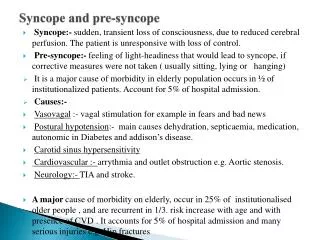
Syncope The current guideline- 2017
What is Syncope? Syncope is the abrupt and transient loss of consciousness associated with absence of postural tone, followed by complete and usuallyrapid spontaneous recovery. The underlying mechanism is global hypoperfusion of both the cerebral cortices or focal hypoperfusion of the reticular activating system (RAS). • •
What is Syncope? It is important to distinguish Syncope from other • causes of T-LOC (Transient Loss of Consciousness) • • • • Pre-Syncope: lightheadedness without LOC Drop Attack: loss of posture without LOC Coma: LOC without spontaneous recovery Seizure: Tonic-Clonic Movements that start WITH LOC (vs hypoxic myoclonus which can occur with syncope), post- ictal recovery period Hypoglycemia • • • • Hypoxia TIA Cardiac Arrest
Etiologies of Syncope • Pain/Noxious Stimuli • Situational(micturation, cough, Neurocardiogenic / Reflex/ Vasovagal defecation) • Fear • Prolongedstanding / heat exposure Carotid Sinus Hypersensitivity (CSH) • Most Common • Arrhythmia–Tachyor Valve Stenosis HOCM(outflowobstruction) Cardiovascular • Drugs: BP meds, Diuretics,TCAs • AutonomicInsufficiency(Parkinsons, OrthostaticHypotension Shy-Dragger, DM, Adrenal “DAAD “ • Alcohol • Dehydration Insufficiency)
Reflex Syncope • The most frequent mechanism for reflex syncope is a mixed cardioinhibitory response, although an individual patient may have syncopal events characterized principally by vasodepressor, cardioinhibitory, or mixed respons • In one cohort of 111 patients with presumed vasovagal syncope who received an implantable loop recorder and were followed for 3 to 15 months, • 34 percent experienced recurrent syncope. • A correlation between syncope and electrocardiographic changes was found in 84 percent, with the most frequent abnormality (seen in 50 percent) being one or more prolonged asystolic pauses, primarily due to sinus arrest. • Bradycardia (<40 beats per minute) without pauses was seen in 9 percent, while the remaining patients had normal sinus rhythm or sinus tachycardia and probably had a vasodepressor response.
ACC/AHA Guideline 2017 Syncope Initial Evaluation
Risk Assessment COR LOE Recommendations Evaluation of the cause and assessment I B-NR B-NR for the short- and long-term morbidity and mortality risk of syncope are recommended. IIb Use of risk stratification scores may be reasonable in the management of patients with syncope.
Work-up and Risk Stratification • The syncope work-up should determine who is at RISK for a dangerous short-term cardiac event. HIGH • All • • • • patient should get basic Work-up Including History/Physical including Orthostatics Medication Review ECG. If age >40, consider Carotid Sinus Massage to assess for Carotid Sinus Hypersensitivity • CONTRAINDICATED if carotid bruit present or recent TIA/Stroke • + Test = bradycardia, hypotension, transient pause/asystole, or prodrome symptoms patients should then be Risk Stratified • All
Work-up and Risk Stratification Risk Stratification • • High Risk: These patients are at high risk for short term cardiac mortality and need appropriate cardiac work-up as an INPATIENT Evidence of significant heart disease (such as heart failure, low left ventricular ejection fraction, structural abnormality, or previous myocardial infarction). Clinical (eg palpitations) or ECG features suggesting arrhythmia Comorbidities such as severe anemia or electrolyte disturbance. • • • • High Risk Work-Up • Echo, Stress test, and/or Ischemic Evaluation Check for recent Echo and/or TMST before ordering a new one! Consider Posterior Circulation imaging of the brain if suspect Neurological “syncope” • • • Carotid Ultrasound has POOR utility in the workup of Syncope and shouldnotbe ordered routinely.
Work-up and Risk Stratification •Low Risk: Patient’s with no High risk characteristics and/or with highly suspected Vasovagal or Neurocardiogenic Etiology Single Episode: No further workup indicated • Multiple Episodes: Can workup as outpatient • Patient having FREQUENT Episodes: Holter Monitor or Event Monitor Patient having INFREQUENT Episodes: Implantable Loop Recorder • • These patients DO NOT need “ACS Rule Out” or Imaging (including Head CT or Carotid Ultrasound) •
Imaging in the Workup of Syncope So • When do I get Brain Imaging? Neurological Causes of true Syncope are RARE • • • Bilateral Carotid or Basilar Artery Disease Non-convulsive Seizure • Head CT is indicated ONLY if the patient has or experienced focal neurological deficits or they experienced head trauma from the event. Carotid Ultrasound has LOW utility and should NOT be ordered • routinely. • Posterior Circulation evaluation with CTA/MRA or Ultrasound is useful only if Vertibro-basilar insufficiency is suspected • TypicallypresentwithDizziness,gait instability, blurryvision, nystagmus, or frankComa.
Neurological and Imaging Diagnostics- 2017 COR LOE Recommendations IIa C-LD Simultaneous monitoring of an EEG and hemodynamic parameters during tilt-table testing can be useful to distinguish among syncope, pseudosyncope, and epilepsy. III: No Benefit III: No Benefit III: No Benefit B-NR B-NR B-NR MRI and CT of the head are not recommended in the routine evaluation ofpatients with syncope in the absence offocal neurological findings or head injurythatsupport further evaluation. Carotid arteryimaging isnotrecommended in the routine evaluation ofpatientswith syncope inthe absence of focal neurological findings thatsupportfurtherevaluation. Routine recording of an EEG is not recommended in the evaluation of patients with syncope in the absence of specific neurological features suggestive of a seizure.
Cardiovascular Testing Cardiac Imaging COR LOE Recommendations Transthoracic echocardiography can be useful in selected patients presenting with syncope if structural heart disease is suspected. CT or MRI may be useful in selected patients presenting with syncope of suspected cardiac etiology. Routine cardiac imaging is not usefulin the evaluation of patients with syncope unlesscardiac etiologyis suspected on the basis ofan initial evaluation, including history, physical examination,or ECG. IIa B-NR B-NR B-R IIb III: No Benefit Troponin should not be routinely done
Stress Testing COR LOE Recommendation Exercise stresstesting can be usefulto establish the cause ofsyncope in selected patientswho experience syncope or presyncope during exertion. IIa C-LD
In-Hospital Telemetry COR LOE Recommendation Continuous ECGmonitoring isuseful for hospitalized patients admitted for syncope evaluation with suspected cardiac etiology. I B-NR • Depends on access and patient population • Overused
Electrophysiological Study COR LOE Recommendations EPScan be useful for evaluation ofselected patients with syncope ofsuspected arrhythmic etiology. IIa B-NR B-NR III: No Benefit EPS is not recommended for syncope evaluation in patients with a normal ECG and normal cardiac structure and function, unless an arrhythmic etiology is suspected.
Tilt-Table Testing COR LOE Recommendations If the diagnosis is unclear after initial evaluation, IIa IIa IIa IIa B-R B-NR B-NR B-NR B-R testing can be useful for patients with suspected VVS. Tilt-table testing can be useful forpatients with syncope and suspected delayed OH when initialevaluation is not diagnostic. Tilt-table testing is reasonable to distinguish convulsive syncope from epilepsy in selected patients. Tilt-table testing isreasonable toestablish a diagnosisof pseudosyncope. Tilt-table testing is not recommended to predict a response to medical treatments for VVS. III: No Benefit
Key Points Key Differential Dx • Vasovagal/Neurocardiogenic - most common Cardiac – HIGH RISK PATIENTS, most dangerous Orthostatic – “D A A D” Other - Neurologic, Functional, Psych o o o o Work-up and Risk Stratification • H/P, Orthostatics, Meds, ECG, +/- Carotid Massage Risk Stratify o o High Risk - Admit w/ cardiac work-up Low Risk - Outpatient workup based on frequency of episodes Brain Imaging ONLY if focal Neuro Deficits or Head trauma •