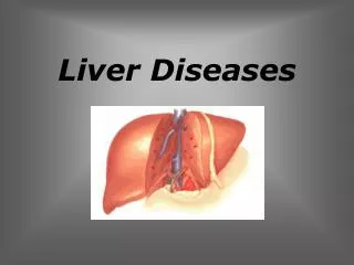
Liver Diseases
Liver Diseases. Benign Liver Tumors. Benign Liver Tumors. Benign liver tumors are relatively common but usually subclinical . Most are detected incidentally by ultrasound (US) or other scanning techniques.

Liver Diseases
E N D
Presentation Transcript
Benign Liver Tumors • Benign liver tumors are relatively common but usually subclinical. • Most are detected incidentally by ultrasound (US) or other scanning techniques. • Others are discovered because of hepatomegaly, right upper quadrant discomfort, or intraperitoneal hemorrhage. • Liver function tests are usually normal The most important benign tumor of the liver are cavernous hemangioma, focal nodular hyperplasia(FNH) and hepatocellular adenoma (HA, liver cell adenoma – LCA).
Cavernous Hemangioma Cavernous hemangioma is the most common primary liver tumor; its occurrence in the general population ranges from 0.4-20%. Cavernous hemangioma is a benign connective tissue tumor resulted from endothelial cells proliferation. It is a non-encapsulated tumor, with an infiltrative, lobular growing. The tumor consists of large (cavernous) spaces, lined by tumor endothelial cells
Cavernous Hemangioma • Usually, they occur as solitary lesions. • However, they may be multiple in as many as 50% of patients. • Hemangiomas typically measure less than 5 cm; some authors call those larger than 4-5 cm giant hemangiomas. Hemangioma showing characteristic sharp demarcation from the surrounding liver and "spongy" texture.
Cavernous Hemangioma • The vast majority of hemangiomas (as many as 85%) are asymptomatic. • Because of advances in imaging technology, hemangiomas are being detected more frequently. • Hemangiomas may cause symptoms because of the compression of adjacent structures, acute thrombosis, or consumptive coagulopathy (Kasabach-Merritt syndrome - giant hemangiomas). • Spontaneous or posttraumatic rupture is a catastrophic complication that occurs in about 1-4% of hemangiomas; it has a considerable mortality rate, as high as 60%
Cavernous Hemangioma • The classic diagnostic findings for hemangioma are as follows: • on unenhanced CT, hypoattenuation similar to that of vessels; • on dynamic contrast-enhanced CT or MR imaging, peripheral globular enhancement anda centripetal fill-in pattern with the attenuation of enhancing areas identical to that of the aorta and blood pool; • on T2- and heavily T2-weighted MR imaging, hyperintensity similar to that of cerebrospinal fluid; • on sonography, homogeneous hyperechogenicity or hypo- or isoechogenicity with a hyperechoic rim; • on delayed phases of 99mTc RBC scanning, a defect in the early phases that shows prolonged and persistent filling-in. From: Gore RM, Levine MS. Textbook of gastrointestinal radiology, 2nd ed. Philadelphia: Saunders, 2000: 1487-1497
Cavernous Hemangioma CT Findings: • Hemangiomas are enhancing lesions that have characteristic dynamic features after the administration of contrast material. • On nonenhanced CT scans, hemangiomas appear hypoattenuating relative to the adjacent liver. • During the arterial-dominant phase, small hemangiomas show intense and uniform contrast enhancement and retain their contrast enhancement during the portal venous phase CT of the liver following intravenous contrast medium administration at 20 sec, at 40 sec, at 1 minute and at 3 minutes, respectively. Early clear enhancement of peripheral vascular lakes is rapidly followed by progressive opacification of the central portions of the haemangioma. At 3 minutes following contrast injection the whole lesion is markedly enhanced relative to the liver.
Cavernous Hemangioma CT Findings: The pattern of a peripheral, discontinuous, intense nodular enhancement during the arterial-dominant phase with progressive centripetal fill-in is considered pathognomic for hemangiomas 30’’ 0’’ 15’ 1’ Contrast-enhanced CT scans reveal the pathognomic features of a hemangioma, namely, peripheral nodular enhancement and progressive centripetal fill-in (arrow).
Cavernous Hemangioma Ultrasound At ultrasonography, hemangiomas appear as well-circumscribed, uniformly hyperechoic lesions
Cavernous Hemangioma Ultrasound • Atypical features include hypoechoic lesions with a thin hyperechoic rim or a thick rind and scalloped borders • Hemangiomas may appear hypoechoic in fatty livers
Cavernous Hemangioma MRI Findings: • MRI is more sensitive and specific than other imaging modalities in the diagnosis of hemangiomas. • Hemangiomas appear as smooth, lobulated, homogeneous, hypointense lesions on T1-weighted images. • On T2-weighted images, they appear hyperintense relative to liver. The high signal intensity on T2-weighted images is due to the extremely long T2 relaxation time of the free fluid (slowly moving blood)
Cavernous Hemangioma MRI Findings: With the injection of contrast material (gadolinium chelates), lesions typically demonstrate peripheral nodular enhancement with progressive, centripetal fill-in that usually appears after 5-30 minutes
Cavernous Hemangioma • Angiography • Angiography has been used extensively in the past as the gold standard to characterize CH but is not applied any more for this purpose due to the several noninvasive imaging methods now available. • It is, however, important to know the angiographic features of this very common hepatic lesion because it may be displayed on hepatic arteriograms performed for other purposes. Angiography of the hepatic artery, arterial phase. Multiple nodular contrast accumulations in C-shaped configuration.
Focal Nodular Hyperplasia Focal nodular hyperplasia (FNH) is the second most common tumor of the liver and constitutes 8% of all liver tumors • FNHoccurs predominantly in women (80 - 90%) during the third to fifth decade. • FNH is not a true neoplasm, and it is believed to represent a hyperplastic response to increased blood flow in an intrahepatic arteriovenous malformation. • Kupffer cells are usually present in the lesion • Most cases of FNH occur as a solitary lesion (80-95%)
Focal Nodular Hyperplasia • FNH is normally a solitary, lobulated, nonencapsulated tumour of relatively small size; usually less than 5 cm in diameter. • It is frequently located in the subcapsular areas of the liver and may be pedunculated. • FNH frequently displays a central fibrous structure or "scar" with multiple radiating fibrous septa. Typically this scar is strongly vascularized by branches coming from the hepatic artery. A classic focal nodular hyperplasia, paler than the surrounding liver, and with a distinct central stellate scar.
Focal Nodular Hyperplasia • In most patients, FNH is discovered incidentally during imaging or laparotomy for unrelated conditions. Most patients are asymptomatic. • A small minority (10-15%) may present with vague abdominal symptoms from mass effect, a palpable mass, or hepatomegaly. • The most common complication of FNH is hemorrhage observed in only 2-3% Although FNH usually has no clinical significance, recognition of the radiological characteristics of FNH is important to avoid unnecessary surgery, biopsy, and follow-up imaging.
Focal Nodular Hyperplasia • The imaging modalities that can best characterize FNH are those that can delineate the central scar or show Kupffer-cell activity: • US, particularly when combined with duplex Doppler US, may be the only type of imaging required. • CT and MRI demonstrate the central scar best, whereas radionuclide scans best demonstrate Kupffer-cell activity. • The introduction of MRI superparamagnetic contrast agents may challenge the role of radionuclide scanning in the future.
Focal Nodular Hyperplasia Ultrasound • For imaging of the right upper quadrant, ultrasonography (US) is more widely used than other modalities, and usually, US findings raise the possibility of FNH. There is a large (diameter 9 cm) area with iso- or slightly hyperechoic pattern in the left liver lobe. The lesion is bordered by a hyperreflective rim.
Focal Nodular Hyperplasia Ultrasound Doppler ultrasound shows centrifugal arterial flow originating from the central portion of the lesion or sometimes from a central vessel with a stellate configuration.
Focal Nodular Hyperplasia • Typical CT finding of a FNH: • In the native scan this benign tumor appears to be almost isodense to the surrounding liver tissue; in its center a "scar" of lower density can be seen distinctly (top left image). • 25 secs after contrast agent application, enhancement starts (top middle image), reaching its maximum after 30-40 secs (top right image). Note the typical delicate structure of the hypodense septae, which appear in a radial order. • After 45-60 secs, homogenous contrasting is reached (bottom left image); this is followed by a gradual decay in contrasting. CT Findings:
Focal Nodular Hyperplasia MRI Findings: • FNH usually has homogeneous signal intensity on MRIs. • The lesion is isointense to hypointense on T1-weighted images in 94-100% of patients. • On T2-weighted images, the lesion is slightly hyperintense to isointense in 94-100% of patients. • The central scar is hypointense on T1-weighted images, but it shows a variable signal-intensity pattern on T2-weighted images.
Focal Nodular Hyperplasia MRI Findings: T1-weighted sequence. A focal lesion (arrow) with a diameter of 2.5 cm is hypointense relative to the liverparenchyma. T2-weighted sequence. The lesion is slightly and inhomogeneously hyperintense.
Focal Nodular Hyperplasia MRI Findings: If a hepatic mass contains a low signal central scar on T1-weighted images that enhances after gadolinium administration, the diagnosis of FNH is fairly certain. Contrast-enhanced T1-weighted MR image, obtained 4 min after injection, shows that lesion remains slightly hyperintense to normal liver, but central scar is highly enhanced (arrowhead).
Focal Nodular Hyperplasia Nuclear Scintigraphy: Hepatobiliary scintigraphy (performed with Tc-99m HIDA or an analogue) is useful in diagnosing focal nodular hyperplasia – it may show normal-to-increased uptake in 40-70% of patients. "hot spot" In the late phase, a large area of increased residual activity marks the FNH in the right lobe. In the early phase, the activity is low.
Hepatocellular Adenoma • Hepatocellular adenoma (HA) is a rare benign tumor of the liver. Two types of HAs have been identified, including tumors of bile duct origin and tumors of liver cell origin: • HAs of bile duct origin usually are smaller than 1 cm and not of clinical interest; typically, they are found incidentally on postmortem examinations. • HAs of liver origin are larger and often are clinically significant. On average, they measure 8-15 cm.
Hepatocellular Adenoma • HA is the most important benign tumor of the liver. • Although HAs may be idiopathic, the lesions most often are seen in young women using oral contraceptives. • (The incidence among long-term users of oral contraceptives is approximately 4 cases per 100,000. In women who do not use oral contraceptives or have used them for less than 2 years, the incidence is 1 case per million) • HAs may rupture and bleed, causing right upper quadrant pain. • (Rarely, rupture may lead to hemorrhagic shock) • HA may undergo malignant degeneration. • (Even at histopathological study it may be difficult to differentiate adenoma from well differentiated hepatocellular carcinoma) Surgical resection is advocated in most patients with presumed HAs.
Hepatocellular Adenoma • Most adenomas are not specifically diagnosed at US and are usually further evaluated with CT or other imaging modalities. • Color Doppler US may help differentiate HA from FHN. • Multiphasic helical CT allows more accurate detection and characterization of focal hepatic lesions. • HAs are typically bright on T1-weighted magnetic resonance images and predominantly hyperintense relative to liver on T2-weighted images. • Findings at radionuclide scintigraphy are rarely diagnostic for HA • Understanding the imaging appearance of HAs can help avoid misdiagnosis and facilitate prompt, effective treatment. From: Luigi Grazioli et al. Hepatic Adenomas: Imaging and Pathologic FindingsRadiographics. 2001;21:877-892.
Hepatocellular Adenoma Ultrasound • On US, HAs demonstrate variable echogenicity. • The most adenomas are not specifically diagnosed at US and are usually further evaluated with CT or other imaging modalities. Sagittal US scan of the liver shows a well-defined, homogeneous, hyperechoic lesion in the right lobe (arrow). Transverse US scan of the liver shows a hypoechoic lesion (cursors) with a hyperechoic center (arrow) due to recent hemorrhage. From: Luigi Grazioli et al. Hepatic Adenomas: Imaging and Pathologic FindingsRadiographics. 2001;21:877-892.
Hepatocellular Adenoma Ultrasound • Color Doppler US may demonstrate: • peripheral peritumoral vessels and • intratumoral vessels that typically have a flat continuous waveform. From: Luigi Grazioli et al. Hepatic Adenomas: Imaging and Pathologic FindingsRadiographics. 2001;21:877-892.
Hepatocellular Adenoma CT Findings: A multiphasic CT scan should be performed to better characterize most hepatic tumors. • On CT, the most consistent finding in HAs is the enhancement pattern. Most lesions show homogeneous enhancement in the hepatic arterial phase.(Unfortunately, this feature is not specific to HAs, since HCC, hypervascular metastases, and FNH can demonstrate similar enhancement in the hepatic arterial phase.) • Since HAs are composed histologically of uniform hepatocytes, most are isoattenuating to healthy liver tissue on nonenhanced scans in the portal venous phase. • The finding of hemorrhage as an area of high attenuation can be seen in as many as 40% of patients. • Typically, HAs have well-defined borders and do not have lobulated contours. • A low-attenuation pseudocapsule can be seen in as many as 25% of patients.
Hepatocellular Adenoma CT Findings: Arterial-phase CT scan shows multiple hypervascular lesions (arrows). On a portal venous-phase CT scan, the adenomas are isoattenuating relative to the surrounding parenchyma. From: Luigi Grazioli et al. Hepatic Adenomas: Imaging and Pathologic FindingsRadiographics. 2001;21:877-892.
Hepatocellular Adenoma CT Findings: Unenhanced CT scan shows a hypoattenuating lesion with high-attenuation blood centrally (arrow). From: Luigi Grazioli et al. Hepatic Adenomas: Imaging and Pathologic FindingsRadiographics. 2001;21:877-892.
Hepatocellular Adenoma MRI Findings: • Some MRI findings are similar to CT findings; however, MRI usually is more sensitive in detecting fat and hemorrhage. • HAs tend to be hyperintense or isointense to liver tissue on T1-weighted images • (Other hepatic lesions can be hyperintense on T1-weighted images, such as melanoma metastases and cavities containing proteinaceous material) • On T2-weighted images, HAs most often are slightly hyperintense to liver tissue. • (This finding is not specific since many hepatic lesions, including HCC and metastases, are hyperintense on T2-weighted images) • After gadolinium administration, the pattern of enhancement is similar to that of CT. • (Most HAs show intense enhancement in the arterial phase and are isointense to liver tissue on delayed imaging) • A central scar has never been reported in an HA.
Hepatocellular Adenoma MRI Findings: a b (a) Portal venous-phase CT scan shows a poorly enhancing, spheric mass without obvious hemorrhage. (b) On a T2-weighted MR image, the mass appears heterogeneously hyperintense. (c) T1-weighted MR image shows the mass with heterogeneous hyperintensity due to hemorrhage. c From: Luigi Grazioli et al. Hepatic Adenomas: Imaging and Pathologic FindingsRadiographics. 2001;21:877-892.
Hepatocellular Adenoma MRI Findings: Large HCC with a mosaic pattern, a tumor capsule, and fatty infiltration. Axial fat-saturated delayed contrast-enhanced MR image shows enhancement of a tumor capsule (arrow). On routine MRI of the liver consisting of T1-weighted and T2-weighted images, chemical-shift imaging, and dynamic gadolinium-enhanced imaging, distinguishing between HAs, HCC, and hypervascular metastases usually is not possible.
Hepatic Cyst • The term hepatic cyst usually refers to solitary nonparasitic cysts of the liver, also known as simple cysts. Most patients with simple cysts are asymptomatic and require no treatment. • The precise frequency of liver cysts is not known because most do not cause symptoms, but liver cysts have been estimated to occur in 5% of the population. • The cause of simple liver cysts is unknown, but cysts are believed to be congenital in origin. • Hepatic cysts are not neoplasms. • (The pathophysiology of simple hepatic cysts is related to fluid secretion by the epithelial lining. Typically, the fluid within the cyst has an electrolyte composition that mimics plasma) The clinician has a number of options for imaging the liver in patients with hepatic cysts. A practical problem in the evaluation of a patient with a cystic hepatic lesion is differentiating cystic neoplasms from simple cysts.
Hepatic Cyst Simple cysts tend to have homogenous low-density interiors The margin of the cystic lesion is well defined and the cyst wall is thin and smooth.