Download
1 / 16
160 likes | 356 Views
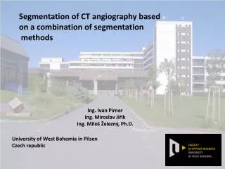
Segmentation of CT a ng iography based on a combination of segmentation methods. Ing. Ivan Pirner Ing. Miroslav Ji řík Ing. Milo š Železný, Ph.D. University of West Bohemia in Pilsen Czech republic. Segmentation of CT angiography Ing . Ivan Pirner. Contents: medical images

E N D
Segmentation of CT angiography based on a combination of segmentation methods Ing. Ivan Pirner Ing. Miroslav Jiřík Ing. Miloš Železný, Ph.D. University of West Bohemia in Pilsen Czech republic
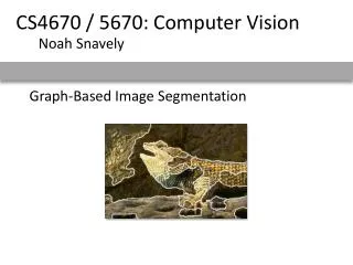
Segmentation of CT angiography Ing. Ivan Pirner Contents: medical images segmentation techniques thresholding edge detection region growing process conclusion Sources of medical images: CT, MRI, USG, X-ray, PET, etc. Example: CT (picture source: http://www.jnch.nic.in) 2 of 16 PRIA 2010, Saint Peterburg 8th December
Segmentation of CT angiography Ing. Ivan Pirner Contents: medical images segmentation techniques thresholding edge detection region growing process conclusion Image properities: Image F(i,j) is a 2-dimensional array of pixels. Each pixel on the position i,j is characterized by its value – the density. The density is a non-negative integer value belonging to a known finite range, usually 8 or 16bit. Remark: Most of the medical images are grayscale. Methods used in this work may be generalized for color images. 3 of 16 PRIA 2010, Saint Peterburg 8th December
Segmentation of CT angiography Ing. Ivan Pirner Contents: medical images segmentation techniques thresholding edge detection region growing process conclusion Definition: Segmentation = labeling the pixels of an image in such way, that the labels have a strong correlation with real objects observed in the image. Purposes: removing unwanted regions of data counting regions measuring regions 4 of 16 PRIA 2010, Saint Peterburg 8th December
Segmentation of CT angiography Ing. Ivan Pirner Contents: medical images segmentation techniques thresholding edge detection region growing process conclusion Useful segmentation techniques: thresholding (threshold value?) edge-based methods edge image thresholding (threshold value?) region-based methods region growing (homogeneity rule?) graph cut segmentation energy minimization (model?, parameters?) 5 of 16 PRIA 2010, Saint Peterburg 8th December
Segmentation of CT angiography Ing. Ivan Pirner Contents: medical images segmentation techniques thresholding edge detection region growing process conclusion Thresholding: Thresholded image is a binary image G(i,j), where G(i,j) = 1 if F(i,j) > T and G(i,j) = 0 otherwise i, j – spatial coordinates F(i,j) – original image pixels Conditions of use: Object to be segmented has other pixel values range than its background. The output segmentation needs often postprocessing, many dummy segments due to image noise. The threshold value must be chosen properly. 6 of 16 PRIA 2010, Saint Peterburg 8th December
Segmentation of CT angiography Ing. Ivan Pirner Contents: medical images segmentation techniques thresholding edge detection region growing process conclusion Example: left: original CT slice right: double thresholded image 7 of 16 PRIA 2010, Saint Peterburg 8th December
Segmentation of CT angiography Ing. Ivan Pirner Contents: medical images segmentation techniques thresholding edge detection region growing process conclusion Edge detection: The purpose is to find places in the image with significant discontinuities in the image function (big differences in values between neighborning pixels). There are many similar operators, which approximate the first derrivative of the image function. We used Sobel’s operator. first two of four Sobels’ operators (the basic mask is rotating) 8 of 16 PRIA 2010, Saint Peterburg 8th December
Segmentation of CT angiography Ing. Ivan Pirner Contents: medical images segmentation techniques thresholding edge detection region growing process conclusion Proceeding: The edge image is set as an output of a 2D-correlation between the mask and the original image. Results for different directions are summed and the output image then thresholded into a binary image. Conditions of use: The seeked region must be bordered by a “sharp” edge. The threshold value must be chosen properly. 9 of 16 PRIA 2010, Saint Peterburg 8th December
Segmentation of CT angiography Ing. Ivan Pirner Contents: medical images segmentation techniques thresholding edge detection region growing process conclusion Example: left: original CT slice right: edge image 10 of 16 PRIA 2010, Saint Peterburg 8th December
Segmentation of CT angiography Ing. Ivan Pirner Contents: medical images segmentation techniques thresholding edge detection region growing process conclusion Region growing: In this part we used the modified confidence connected algorithm: Set a seed (1 or multiple points) and make it the current region. Find all pixels neighboring upon the current region. For all of this neighboring pixels decide, whether they fulfill the homogeneity criteria, if yes, append them to the current region. If no points added in step 3, END, else GOTO 2. We chose as homogeneity criteria K(p) a double inequality: K(p) = 1 if p>T_min && p<T_max K(p) = 0 otherwise p – tested pixel T_min – chosen minimum value T_max – chosen maximum value 11 of 16 PRIA 2010, Saint Peterburg 8th December
Segmentation of CT angiography Ing. Ivan Pirner Contents: medical images segmentation techniques thresholding edge detection region growing process conclusion Sketch: region growing (image source: http://www.cs.cf.ac.uk) Conditions of use: Seeked region must be homogenous. The seed set must be chosen within the region. 12 of 16 PRIA 2010, Saint Peterburg 8th December
Segmentation of CT angiography Ing. Ivan Pirner Contents: medical images segmentation techniques thresholding edge detection region growing process conclusion Proceeding: original image region growing edge detection segmented bones+vessels edge image morphological operations bone image subtraction segmented vessels 13 of 16 PRIA 2010, Saint Peterburg 8th December
Segmentation of CT angiography Ing. Ivan Pirner Contents: medical images segmentation techniques thresholding edge detection region growing process conclusion Visualization of the 3D data: 3D model using volume rendering: 14 of 16 PRIA 2010, Saint Peterburg 8th December
Segmentation of CT angiography Ing. Ivan Pirner Contents: medical images segmentation techniques thresholding edge detection region growing process conclusion Conclusion: The CT angiography vessel segmentation may be made using “simple” methods when combining them together. Parameters of each of the used segmentation methods can be easily interpreted and either directly determined or experimentally measured. Future work: Graph cuts could bring more precise results, although we need to determine a proper model and estimate its parameters. 15 of 16 PRIA 2010, Saint Peterburg 8th December
Segmentation of CT angiography Ing. Ivan Pirner Contents: medical images segmentation techniques thresholding edge detection region growing process conclusion Thank you for your attention. 16 of 16 PRIA 2010, Saint Peterburg 8th December