Download
1 / 29
290 likes | 608 Views
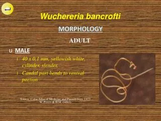
Source: Color Atlas of Medicine and Parasitology. 1977 W. Peters & H.M. Gillers. Wucher er ia bancrofti. MORPHOLOGY. ADULT. MALE 40 x 0,1 mm, yellowish white, cylinder, slender, Caudal part bends to ventral portion.

E N D
Source: Color Atlas of Medicine and Parasitology. 1977 W. Peters & H.M. Gillers Wuchereriabancrofti MORPHOLOGY ADULT • MALE • 40 x 0,1 mm, yellowish white, cylinder, slender, • Caudal part bends to ventral portion
Sumber : Color Atlas of Medicine and Parasitology. 1977. W. Peters & H.M. Gillers Wuchereria bancrofti MORPHOLOGY ADULT • FEMALE • (80-100) x (0,24-0,30) mm, soft, blunt nail, slight rounded head.
Cephalic space Sheath Sheath Slender tail Nuclei MORPHOLOGY OF MICROFILARIAE Source : Medical Parasitology in Plates. G. Piekarski
Microfilaria Brugia malayi Microfilaria Wuchereria bancrofti Microfilaria Brugia timori
Cephalic space (L:W=1:1) Tail (No nuclei) Source : Atlas of Medical Parasitology. Prayong Radomyos, dkk. Pewarnaan hematoxylin Source : Color Atlas of Medicine and Parasitology. 1977 W. Peters & H.M. Gillers. Pewarnaan giemsa Wucheria bancrofti MORPHOLOGY MICROFILARIAE
Cephalic space (L:W=2:1) B. malayi W. bancrofti Bloated tail (nucleus in it) Brugia malayi MORPHOLOGY MICROFILARIAE
Source : Atlas of Medical Parasitology. Prayong Radomyos, dkk. Brugia timori MORPHOLOGY MICROFILARIAE • Looking like microfilariae B. malayi • Size 287-341 • Sheathed not clear at coloration of Giemsa (dr. Djaenudin Natadisastra, SpParK)
Source : Atlas of Medical Parasitology. Prayong Radomyos, dkk. Brugia timori MORPHOLOGY MICROFILARIAE • Stiff body broken, tip of tail rather blunt • “Nuclei” in the body, spread in the irregular groups, presence of (5-8) nuclei in the tail • Smaller nuclei at the distal than in B. malayi • Cephalic space: 3 : 1
GENERAL MORPHOLOGY Plasmodium sp. IN ERYTHOCYTIC CYCLES • FOUND TWO MAIN STAGE : • TROPHOZOITE STAGE • Ring form • Old/Matured trophozoite • SCHIZONT STAGE • Young schizont • Old/Matured schizont
Protoplasm Nucleus ERYTHROCYTE • Ring Form with reddish Nucleus and bluish Protoplasm • Chromatin dots Plasmodium sp. MORPHOLOGY TROPHOZOITE STAGE
Protoplasm Nucleus ERYTHROCYTE Nucleus and Protoplasm grow bigger Plasmodium sp. MORPHOLOGY TROPHOZOITE STAGE
Protoplasm Nucleus ERYTHROCYTE • Nucleus starts fission process • Protoplasm remains in size Plasmodium sp. MORPHOLOGY SCHIZONT STAGE
Merozoite ERYTHROCYTE • Complete Merozoites • Note the Regularity, Pigment Plasmodium sp. MORPHOLOGY SCHIZONT STAGE
ERYHTROCYTE Ring Form ERYTHROCYTE • Ring Form • Erythrocyte enlarged, pale • Schuffner dots PLASMODIUM VIVAX TROPHOZOITE STAGE
ERYTHROCYTE ERYTHROCYTE Amoeboid Form Protoplasm grows (Amoeboid form) with vacuole PLASMODIUM VIVAX TROPHOZOITE STAGE Vacuole
Fission of Nucleus ERYTHROCYTE • Fission of Nucleus • Vacuole disappear PLASMODIUM VIVAX SCHIZONT STAGE
Pigment MEROZOITe • Merozoite (12-14) • Pigment PLASMODIUM VIVAX SCHIZONT STAGE
Pigment NUCLEUS NUCLEUS • Round/Oval • Small nucleus , compact, eccentric • Bluish protoplasm • Round/Oval • Big nucleus, pale • Red-blue protoplasm PLASMODIUM VIVAX GAMOTOCYTE STAGE: Macro and Microgametocyte
PLASMODIUM OVALE TROPHOZOITE STAGE • Ring Form (thick) • Erythrocyte • Chromatin dots
PLASMODIUM OVALE TROPHOZOITE STAGE Round, compact trophozoite Erythrocyte deformed Schuffner/James’ Dots
Resemble P. vivax Normal erythrocyte Plasmodium malariae TROPHOZOITE STAGE
Trophozoite Band form Trophozoite Plasmodium malariae TROPHOZOITE STAGE
Pigment MEROZOITE • Rossete-like • Merozoit (6-12) Plasmodium malariae SCHIZONT STAGE
Round/Oval • Large nucleus, pale • Reddish protoplasm • Round/Oval • Small nucleus, compact • Bluish protoplasm Plasmodium malariae GAMETOCYTE STAGE: Macro and Microgametocyte
TROPHOZOITE STAGE Plasmodium falciparum Fine ring Multiple infection
Vacuole • Vacuole surrounded by plasma • Maurer’s dots Plasmodium falciparum TROPHOZOITE STAGE
Schizont occupy 2/3 erythrocyte • 8-24 Merozoite Plasmodium falciparum SCHIZONT STAGE
Plasmodium falciparum GAMETOCYTE STAGE: Macro and Microgametocyte • Sausage shape • Large nucleus, pale, more eccentric position • Pigment dispersed • Reddish protoplasm • Banana/crescent shape • Small nucleus, compact, in the middle • Pigment around nucleus • Bluish protoplasm
THICK BLOOD PREPARATION Plasmodium falciparum PLASMODIUM VIVAX • Uniform • No red zone around parasite • Smaller than lymphocyte nucleus Plasmodium malariae • Not uniform • No red zone around parasite • Smaller than lymphocyte nucleus • Not uniform • Red zone around parasite • Bigger than lymphocyte nucleus