Download
1 / 14
140 likes | 278 Views
Fetal Studio offers affordable and professional access to prenatal ultrasound, limited medical surveys and level 1 ultrasounds, <br>also the best 3D & 4D imaging quality available on the market.

E N D
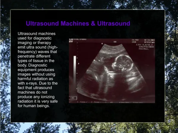
Ultrasound Machines & Ultrasound Ultrasound Machines & Ultrasound Ultrasound machines used for diagnostic imaging or therapy emit ultra sound (high- frequency) waves that penetrate different types of tissue in the body. Diagnostic equipment produces images without using harmful radiation as with x-rays. Due to the fact that ultrasound machines do not produce any ionizing radiation it is very safe for human beings.
Ultrasound Machines & Ultrasound With their own easily obtainable ultrasound machines, doctors' offices and clinics are able to examine various organs in the body on-site without having to send patients to the hospital or expensive medical imaging centers. Compared to magnetic resonance imaging (MRI) and computed tomography (CT), ultrasonography is relatively inexpensive and the equipment is very portable. With today's healthcare environment ultrasound can be considered the most cost effective modern imaging modality.
What is Ultra Sound? Ultra- is used to describe sound waves as in "ultrasound", that which exceeds a frequency that a human ear can detect. Normally a human ear can hear approximately 20 hertz to 20,000 hertz (1 hertz is one cycle per second). Ultra sound, used for diagnostic imaging or therapy, can be employed to analyze or treat medical conditions. It applies cyclical sound pressure pulsed at a frequency higher than the human ear can detect, or 20,000 hertz, hence the name ultrasound.
Components of an Ultrasound Exam Sonographers & Ultrasound Technologists are trained operators of ultrasound machines Central Processing Unit (CPU) - The ultrasound machine's brain. The computer that contains the microprocessor, memory, amplifiers and power supplies for the microprocessor and transducer probe. The CPU sends electrical currents to the transducer, which in turn sends electrical pulses that bounce off of the target and return echoes. The CPU calculates the location of thousands of points of echo origins to produce an image for output such as a monitor, a printer, a network drive; or a disk.
Transducer Pulse Controls The transducer pulse controls can set and vary the frequency, amplitude and duration of the ultra sound pulses. Electrical currents are applied to the piezoelectric (PZ) crystals in the transducer/ probe^, as selected by the operator. Display - The display turns processed data from the CPU into an image. Sonogram images have typically been black-and-white, but newer ultrasound machines can produce color Doppler images.
Transducer Pulse Controls Display - The display turns processed data from the CPU into an image. Sonogram images have typically been black-and-white, but newer ultrasound machines can produce color Doppler images. Keyboard/Cursor - Ultrasound machines have a keyboard and a cursor. The keyboard allows the operator to add notes and to take measurements of the image; the mouse enables the operator to interact with the ultrasound machines software.
Disk Storage - The processed data and/or images can be stored. Storage can include hard disks, compact disks (CDs), digital video disks (DVDs), or a network drive. Most of the time, ultrasound machines store data with the patient's medical records. Ultrasound Printers - Most ultrasound machines have thermal or digital printers connected to them. Ultrasound images are in motion (real time), but a still/ frozen image can be captured at any point and sent to the printer for printing.
Ultrasound Uses Ultra sound machines can be used on many objects, typically to penetrate them and evaluate the reflections that occur, or to supply concentrated energy. The ultra sound reflection signature can detail the internal structure of the medium. The most well known exam performed on ultrasound machines is the obstetric ultrasound exam (imaging of the fetus). While many people relate this applied science with obstetrics only, there are many other applications for ultrasound technology. Now a days ultrasound is performed in small offices and private clinics. Within the last decade many endocrinologists and otolaryngologists have turned to performing ultrasound exams in the comfort of their own offices. Identifying and diagnosing many ailments using ultrasound imaging has steeply declined our death rate from a variety of diseases and conditions. Medical diagnosis, which is crucial in disease treatment, has been a chief cause in the overall improvement of healthcare outcomes. Ultrasound machines have given a huge boost to medical diagnosis. Ultrasound machines can visualize problems in patients' organs and other tissues by bouncing ultra sound waves off of them and using a computer to plot the many points received and portrays these plots into images on a monitor or print them onto thermal paper.
Ultrasound Uses Ultra sound machines can be used on many objects, typically to penetrate them and evaluate the reflections that occur, or to supply concentrated energy. The ultra sound reflection signature can detail the internal structure of the medium. The most well known exam performed on ultrasound machines is the obstetric ultrasound exam (imaging of the fetus). While many people relate this applied science with obstetrics only, there are many other applications for ultrasound technology. Now a days ultrasound is performed in small offices and private clinics. Within the last decade many endocrinologists and otolaryngologists have turned to performing ultrasound exams in the comfort of their own offices. Identifying and diagnosing many ailments using ultrasound imaging has steeply declined our death rate from a variety of diseases and conditions. Medical diagnosis, which is crucial in disease treatment, has been a chief cause in the overall improvement of healthcare outcomes. Ultrasound machines have given a huge boost to medical diagnosis. Ultrasound machines can visualize problems in patients' organs and other tissues by bouncing ultra sound waves off of them and using a computer to plot the many points received and portrays these plots into images on a monitor or print them onto thermal paper.
Diagnostics - Capture size, structure, and any pathological lesions; Scans routinely conducted are obstetric, cardiac, renal, hepatic (liver) and gallbladder. Other applications include musculo-skeletal (imaging of muscles, ligaments and tendons), endocrinology and otolaryngology (thyroid, salivary glands, and lymph nodes), ophthalmic ultrasound (eye) scans and other superficial structures such as breast and testicles. Ultrasound is also increasingly being used in trauma and first aid cases by (the likes of) EMT response teams. Because of the real time nature of ultrasound, it is often used to guide interventional procedures such as fine needle aspiration FNA or biopsy of masses for cytology or histology testing in the breast, thyroid, liver, kidney, lymph nodes. Vascular scans are possible with the use of Doppler to display blood flow. Doppler is also being used to evaluate the blood flow of organs and cancerous regions.
Therapeutic - pulverize kidney- and gall-stones; focused ultrasound surgery; acoustic targeted drug delivery; cataract treatment; stimulate tooth and bone growth; non-surgical treatment of varicose veins; liposuction and lipectomy; bacterial cell killing; and acoustophoresis (contactless separation, concentration, and manipulation of microparticles and biological cells). Again the real time nature of ultrasound assists in injecting medications into muscles and joints. Industrial Cleaning - teeth and medical / dental instruments Humidifiers Echo Location & Range Finding Chemistry Weaponry
Ultrasound machines use ultra sound These waves are emitted by the machine and bounce back when they collide with normal tissue or tumors. The reflections occur at boundaries between different tissue types. The machine times the reflections to calculate distances and generate images of the organs. The different shades in an ultrasound image are equivalent to the intensities of the reflectors. Ultrasound machines provide the ability to view live images of our internal organs. By using the controls provided, a sonographer can view the exact section of an internal organ. (Portable machines are also available for emergency medical teams.)
How an ultrasound is done? In an ultrasound scan, a real-time scanner forms a continuous range of images of a subject and places it on the monitor. A transducer is used to produce these waves. The recurring ultrasound beams scan the subject and then go back to the transducer after reflection. The data obtained from the different reflections recomposed in the form of a picture that is displayed on a screen. Ultrasound imaging is a complex medical procedure that requires prior training. The potential health risks that generated high frequency waves can produce damaging the body tissue if exposure is too lengthy warrant this. As such, only professional doctors and registered diagnostic medical sonographers with experience in their field of expertise can correctly regulate the duration of an exam.
Is Ultrasound Safe? When applied by trained ultrasound technologists or sonographers, ultrasound machines are generally safe with no known risks to patients because ultra sound machines do not use mutagenic ionizing radiation. If applied at length or at higher than diagnostic power, there potentially could be three effects created by ultrasonic energy: These could be enhanced inflammation; heating of soft tissue from molecular friction; and/or production of microscopic bubbles in living tissues which creates distortion of the cell membrane. This though is not known to occur at diagnostic power levels used by modern diagnostic machines. Ultrasound machines use Ultra sound in a generally safe manner. What makes medical sonography safe is its high frequency and low loudness, lack of radiation, and skillful application by trained sonographers. When it comes to ultrasound, machines of this complexity are only to be used by trained ultrasound technicians or sonogram machine operators who are experts in medical ultrasound. Machines are very expensive and highly advanced.Article Source: http://EzineArticles.com/expert/Steve_ Chard/502913