Download
1 / 86
860 likes | 874 Views
CHAPTER 17 FROM GENE TO PROTEIN. Section A: The Connection Between Genes and Proteins. 1. The study of metabolic defects provided evidence that genes specify proteins 2. Transcription and translation are the two main processing linking gene to protein: an overview

E N D
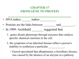
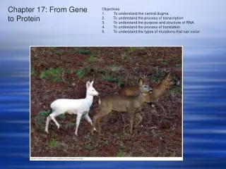
CHAPTER 17 FROM GENE TO PROTEIN Section A: The Connection Between Genes and Proteins 1. The study of metabolic defects provided evidence that genes specify proteins 2. Transcription and translation are the two main processing linking gene to protein: an overview 3. In the genetic code, nucleotide triplets specify amino acids 4. The genetic code must have evolved very early in the history of life
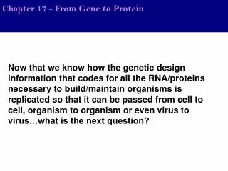
The information content of DNA is in the form of specific sequences of nucleotides along the DNA strands. The DNA inherited by an organism leads to specific traits by dictating the synthesis of proteins. Proteins are the links between genotype and phenotype. For example, Mendel’s dwarf pea plants lack a functioning copy of the gene that specifies the synthesis of a key protein, gibberellins. Gibberellins stimulate the normal elongation of stems. Introduction
In 1909, Archibald Gerrod was the first to suggest that genes dictate phenotype through enzymes that catalyze specific chemical reactions in the cell. The symptoms of an inherited disease reflect a person’s inability to synthesize a particular enzyme. Gerrod speculated that alkaptonuria, a hereditary disease, was caused by the absence of an enzyme that breaks down a specific substrate, alkapton. Research conducted several decades later supported Gerrod’s hypothesis. 1. The study of metabolic defects provided evidence that genes specify proteins
Progress in linking genes and enzymes rested on the growing understanding that cells synthesize and degrade most organic molecules in a series of steps, a metabolic pathway. In the 1930s, George Beadle and Boris Ephrussi speculated that each mutation affecting eye color in Drosophila blocks pigment synthesis at a specific step by preventing production of the enzyme that catalyzes that step. However, neither the chemical reactions nor the enzymes were known at the time.
Beadle and Edward Tatum were finally able to establish the link between genes and enzymes in their exploration of the metabolism of a bread mold, Neurospora crassa. They mutated Neurospora with X-rays and screened the survivors for mutants that differed in their nutritional needs. Wild-type Neurospora can grow on a minimal medium of agar, inorganic salts, glucose, and the vitamin biotin. Most nutritional mutants can survive on a complete growth medium which includes all 20 amino acids.
One type of mutant required only the addition of arginine to the minimal growth medium. Beadle and Tatum concluded that this mutant was defective somewhere in the biochemical pathway that normally synthesizes arginine. They identified three classes of arginine deficient mutants, each apparently lacking a key enzyme at a different step in the synthesis of arginine. They demonstrated this by growing these mutant strains in media that provided different intermediate molecules. Their results provided strong evidence for the onegene - one enzyme hypothesis.
Fig. 17.1 One gene – one enzyme, now one gene - one polypeptide
Later research refined the one gene - one enzyme hypothesis. First, it became clear that not all proteins are enzymes and yet their synthesis depends on specific genes. This tweaked the hypothesis to one gene - one protein. Later research demonstrated that many proteins are composed of several polypeptides, each of which has its own gene. Therefore, Beadle and Tatum’s idea has been restated as the one gene - one polypeptide hypothesis.
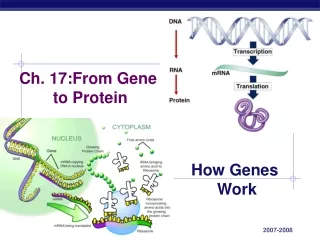
Genes provide the instructions for making specific proteins. The bridge between DNA and protein synthesis is RNA. RNA is chemically similar to DNA, except that it contains ribose as its sugar and substitutes the nitrogenous base uracil for thymine. An RNA molecules almost always consists of a single strand. 2. Transcription and translation are the two main processes linking gene to protein: an overview
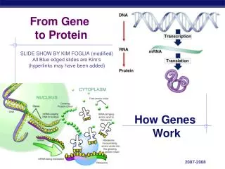
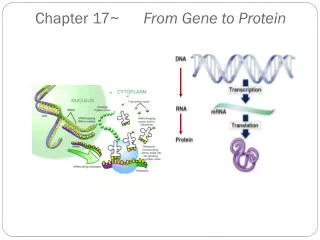
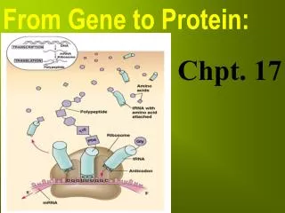
In DNA or RNA, the four nucleotide monomers act like the letters of the alphabet to communicate information. The specific sequence of hundreds or thousands of nucleotides in each gene carries the information for the primary structure of a protein, the linear order of the 20 possible amino acids. To get from DNA, written in one chemical language, to protein, written in another, requires two major stages, transcription and translation.
During transcription, a DNA strand provides a template for the synthesis of a complementary RNA strand. This process is used to synthesize any type of RNA from a DNA template. Transcription of a gene produces a messenger RNA (mRNA) molecule. During translation, the information contained in the order of nucleotides in mRNA is used to determine the amino acid sequence of a polypeptide. Translation occurs at ribosomes.
The basic mechanics of transcription and translation are similar in eukaryotes and prokaryotes. Because bacteria lack nuclei, transcription and translation are coupled. Ribosomes attach to the leading end of a mRNA molecule while transcription is still in progress. Fig. 17.2a
In a eukaryotic cell, almost all transcription occurs in the nucleus and translation occurs mainly at ribosomes in the cytoplasm. In addition, before the primary transcriptcan leave the nucleus it is modified in various ways during RNA processingbefore the finished mRNA is exported to the cytoplasm. Fig. 17.2b
To summarize, genes program protein synthesis via genetic messenger RNA. The molecular chain of command in a cell is : DNA -> RNA -> protein.
If the genetic code consisted of a single nucleotide or even pairs of nucleotides per amino acid, there would not be enough combinations (4 and 16 respectively) to code for all 20 amino acids. Triplets of nucleotide bases are the smallest units of uniform length that can code for all the amino acids. In the triplet code, three consecutive bases specify an amino acid, creating 43 (64) possible code words. The genetic instructions for a polypeptide chain are written in DNA as a series of three-nucleotide words. 3. In the genetic code, nucleotide triplets specify amino acids
During transcription, one DNA strand, the template strand, provides a template for ordering the sequence of nucleotides in an RNA transcript. The complementary RNA molecule is synthesized according to base-pairing rules, except that uracil is the complementary base to adenine. During translation, blocks of three nucleotides, codons, are decoded into a sequence of amino acids. Fig. 17.3
During translation, the codons are read in the 5’->3’ direction along the mRNA. Each codon specifies which one of the 20 amino acids will be incorporated at the corresponding position along a polypeptide. Because codons are base triplets, the number of nucleotides making up a genetic message must be three times the number of amino acids making up the protein product. It would take at least 300 nucleotides to code for a polypeptide that is 100 amino acids long.
The task of matching each codon to its amino acid counterpart began in the early 1960s. Marshall Nirenberg determined the first match, that UUU coded for the amino acid phenylalanine. He created an artificial mRNA molecule entirely of uracil and added it to a test tube mixture of amino acids, ribosomes, and other components for protein synthesis. This “poly(U)” translated into a polypeptide containing a single amino acid, phenyalanine, in a long chain. Other more elaborate techniques were required to decode mixed triplets such a AUA and CGA.
By the mid-1960s the entire code was deciphered. 61 of 64 triplets code for amino acids. The codon AUG not only codes for the amino acid methionine but also indicates the start of translation. Three codons do not indicate amino acids but signal the termination of translation. Fig. 17.4
The genetic code is redundant but not ambiguous. There are typically several different codons that would indicate a specific amino acid. However, any one codon indicates only one amino acid. [If you have a specific codon, you can be sure of the corresponding amino acid, but if you know only the amino acid, there may be several possible codons.] Both GAA and GAG specify glutamate, but no other amino acid. Codons synonymous for the same amino acid often differ only in the third codon position.
To extract the message from the genetic code requires specifying the correct starting point. This establishes the reading frame and subsequent codons are read in groups of three nucleotides. The cell’s protein-synthesizing machinery reads the message as a series of nonoverlapping three-letter words. In summary, genetic information is encoded as a sequence of nonoverlapping base triplets, or codons, each of which is translated into a specific amino acid during protein synthesis.
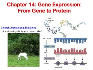
The genetic code is nearly universal, shared by organisms from the simplest bacteria to the most complex plants and animals. In laboratory experiments, genes can be transcribed and translated after they are transplanted from one species to another. This tobacco plant is expressinga transpired firefly gene. 4. The genetic code must have evolved very early in the history of life Fig. 17.5
This has permitted bacteria to be programmed to synthesize certain human proteins after insertion of the appropriate human genes. This and other similar applications are exciting developments in biotechnology. Exceptions to the universality of the genetic code exist in translation systems where a few codons differ from standard ones. These occur in certain single-celled eukaryotes like Paramecium. Other examples include translation in certain mitochondria and chloroplasts.
The near universality of the genetic code must have been operating very early in the history of life. A shared genetic vocabulary is a reminder of the kinship that bonds all life on Earth.
CHAPTER 17 FROM GENE TO PROTEIN Section B: The Synthesis and Processing of RNA 1. Transcription is the DNA-directed synthesis of RNA: a closer look 2. Eukaryotic cells modify RNA after transcription
Messenger RNA is transcribed from the template strand of a gene. RNA polymerase separates the DNA strands at the appropriate point and bonds the RNA nucleotides as they base-pair along the DNA template. Like DNA polymerases, RNA polymerases can add nucleotides only to the 3’ end of the growing polymer. Genes are read 3’->5’, creating a 5’->3’ RNA molecule. 1. Transcription is the DNA-directed synthesis of RNA: a closer look
Specific sequences of nucleotides along the DNA mark where gene transcription begins and ends. RNA polymerase attaches and initiates transcription at the promotor, “upstream” of the information contained in the gene, the transcription unit. The terminator signals the end of transcription. Bacteria have a single type of RNA polymerase that synthesizes all RNA molecules. In contrast, eukaryotes have three RNA polymerases (I, II, and III) in their nuclei. RNA polymerase II is used for mRNA synthesis.
Transcriptioncan beseparatedinto threestages:initiation, elongation, andtermination. Fig. 17.6a
The presence of a promotor sequence determines which strand of the DNA helix is the template. Within the promotor is the starting point for the transcription of a gene. The promotor also includes a binding site for RNA polymerase several dozen nucleotides upstream of the start point. In prokaryotes, RNA polymerase can recognize and bind directly to the promotor region.
STEP 1 - INITIATION In eukaryotes, proteins called transcription factors recognize the promoter region, especially a TATA box, and bind to the promoter. After they have boundto the promoter,RNA polymerasebinds to transcriptionfactors to create atranscriptioninitiation complex. RNA polymerasethen startstranscription. Fig. 17.7
STEP 2 - ELONGATION As RNA polymerase moves along the DNA, it untwists the double helix, 10 to 20 bases at time. The enzyme addsnucleotides to the3’ end of thegrowing strand. Behind the pointof RNA synthesis,the double helixre-forms and theRNA moleculepeels away. Fig. 17.6b
A single gene can be transcribed simultaneously by several RNA polymerases at a time. A growing strand of RNA trails off from each polymerase. The length of each new strand reflects how far along the template the enzyme has traveled from the start point. The congregation of many polymerase molecules simultaneously transcribing a single gene increases the amount of mRNA transcribed from it. This helps the cell make the encoded protein in large amounts.
STEP 3 - TERMINATION Transcription proceeds until after the RNA polymerase transcribes a terminator sequence in the DNA. In prokaryotes, RNA polymerase stops transcription right at the end of the terminator. Both the RNA and DNA is then released. In eukaryotes, the polymerase continues for hundreds of nucleotides past the terminator sequence, AAUAAA. At a point about 10 to 35 nucleotides past this sequence, the pre-mRNA is cut from the enzyme.
Enzymes in the eukaryotic nucleus modify pre-mRNA before the genetic messages are dispatched to the cytoplasm. At the 5’ end of the pre-mRNA molecule, a modified form of guanine is added, the 5’ cap. This helps protect mRNA from hydrolytic enzymes. It also functions as an “attach here” signal for ribosomes. 2. Eukaryotic cells modify RNA after transcription
At the 3’ end, an enzyme adds 50 to 250 adenine nucleotides, the poly(A) tail. In addition to inhibiting hydrolysis and facilitating ribosome attachment, the poly(A) tail also seems to facilitate the export of mRNA from the nucleus. The mRNA molecule also includes nontranslated leader and trailer segments. Fig. 17.8
The most remarkable stage of RNA processing occurs during the removal of a large portion of the RNA molecule during RNA splicing. Most eukaryotic genes and their RNA transcripts have long noncoding stretches of nucleotides. Noncoding segments, introns, lie between coding regions. The final mRNA transcript includes coding regions, exons, that are translated into amino acid sequences, plus the leader and trailer sequences.
Fig. 17.9 • RNA splicing removes introns and joins exons to create an mRNA molecule with a continuous coding sequence.
This splicing is accomplished by a spliceosome. spliceosomes consist of a variety of proteins and several small nuclear ribonucleoproteins (snRNPs). Each snRNP has several protein molecules and a small nuclear RNA molecule (snRNA). Each is about 150 nucleotides long.
(1) Pre-mRNA combineswith snRNPs and otherproteins to form aspliceosome. (2) Within the spliceosome, snRNA base-pairs withnucleotides at the ends ofthe intron. (3) The RNA transcript is cut to release the intron, and the exons are spliced together; the spliceosome then comes apart, releasing mRNA, which now contains only exons. Fig. 17.10
In this process, the snRNA acts as a ribozyme, an RNA molecule that functions as an enzyme. • Like pre-mRNA, other kinds of primary transcripts may also be spliced, but by diverse mechanisms that do not involve spliceosomes. • In a few cases, intron RNA can catalyze its own excision without proteins or extra RNA molecules. • The discovery of ribozymes rendered obsolete the statement, “All biological catalysts are proteins.”
RNA splicing appears to have several functions. First, at least some introns contain sequences that control gene activity in some way. Splicing itself may regulate the passage of mRNA from the nucleus to the cytoplasm. One clear benefit of split genes is to enable a one gene to encode for more than one polypeptide. Alternative RNA splicing gives rise to two or more different polypeptides, depending on which segments are treated as exons. Early results of the Human Genome Project indicate that this phenomenon may be common in humans.
Split genes may also facilitate the evolution of new proteins. Proteins often have amodular architecturewith discrete structuraland functional regionscalled domains. In many cases, different exons code for different domains of a protein. Fig. 17.11
The presence of introns increases the probability of potentially beneficial crossing over between genes. Introns increase the opportunity for recombination between two alleles of a gene. This raises the probability that a crossover will switch one version of an exon for another version found on the homologous chromosome. There may also be occasional mixing and matching of exons between completely different genes. Either way, exon shuffling could lead to new proteins through novel combinations of functions.
CHAPTER 17 FROM GENE TO PROTEIN Section C: The Synthesis of Protein 1. Translation is the RNA-directed synthesis of a polypeptide: a closer look 2. Signal peptides target some eukaryotic polypeptides to specific destinations in the cell 3. RNA plays multiple roles in the cell: a review 4. Comparing protein synthesis in prokaryotes and eukaryotes: a review 5. Point mutations can affect protein structure and function 6. What is a gene? revisiting the question
In the process of translation, a cell interprets a series of codons along a mRNA molecule. Transfer RNA (tRNA) transfers amino acids from the cytoplasm’s pool to a ribosome. The ribosome adds each amino acid carried by tRNA to the growing end of the polypeptide chain. 1. Translations is the RNA-directed synthesis of a polypeptide: a closer look Fig. 17.12
During translation, each type of tRNA links a mRNA codon with the appropriate amino acid. Each tRNA arriving at the ribosome carries a specific amino acid at one end and has a specific nucleotide triplet, an anticodon, at the other. The anticodon base-pairs with a complementary codon on mRNA. If the codon on mRNA is UUU, a tRNA with an AAA anticodon and carrying phenyalanine will bind to it. Codon by codon, tRNAs deposit amino acids in the prescribed order and the ribosome joins them into a polypeptide chain.
Like other types of RNA, tRNA molecules are transcribed from DNA templates in the nucleus. Once it reaches the cytoplasm, each tRNA is used repeatedly to pick up its designated amino acid in the cytosol, to deposit the amino acid at the ribosome, and to return to the cytosol to pick up another copy of that amino acid.
A tRNA molecule consists of a strand of about 80 nucleotides that folds back on itself to form a three-dimensional structure. It includes a loop containing the anticodon and an attachment site at the 3’ end for an amino acid. Fig. 17.13
If each anticodon had to be a perfect match to each codon, we would expect to find 61 types of tRNA, but the actual number is about 45. The anticodons of some tRNAs recognize more than one codon. This is possible because the rules for base pairing between the third base of the codon and anticodon are relaxed (called wobble). At the wobble position, U on the anticodon can bind with A or G in the third position of a codon. Some tRNA anticodons include a modified form of adenine, inosine, which can hydrogen bond with U, C, or A on the codon.
Each amino acid is joined to the correct tRNA by aminoacyl-tRNA synthetase. The 20 different synthetases match the 20 different amino acids. Each has active sites for only a specific tRNA and amino acid combination. The synthetase catalyzes a covalent bond between them, forming aminoacyl-tRNA or activated amino acid. Fig. 17.14