Download
1 / 25

E N D
1. Eukaryotic Genome Organization and Regulation
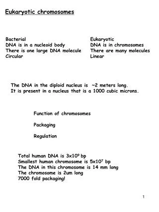
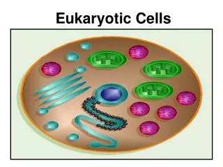
2. Chromatin Organization Each human chromosome averages about 1.5 � 108 nucleotide pairs
Each extended DNA molecule would be about 4 cm long � 1000X longer than the cell diameter
(-) charged DNA wrapped around (+) charged histone proteins
Extreme coiling shortens length of molecule by 100,000X
3. Histone Proteins Among eukaryotic organisms histone proteins are very similar
What might this indicate?
HINT: think about evolution
DNA wrapped around histone proteins � nucleosome
Two molecules each of four types of histone: H2A, H2B, H3, and H4
One molecule of H1 attaches to the DNA near the nucleosome
The amino acid (N-terminus) of each histone protein (the histone tail) extends outward from the nucleosome.
Histones leave the DNA briefly during DNA replication; but not during transcription
4. Why Causes the Coiling? Interactions between H1 + tails of nucleosome + linker DNA on either side cause formation of 30-nm chromatin fiber
Fiber forms looped domains which attach protein scaffold
Particular genes located in the same places on metaphase chromosomes in eukaryotic species � involved in coiling
Interphase chromosomes:
Highly condensed areas (heterochromatin) interspersed with less compact areas (euchromatin)
Heterochromatin DNA cannot be transcribed
The chromatin of each chromosome occupies a specific restricted area within the interphase nucleus � there is an order among the chaos!
Looped domains attached to nuclear envelope in order to secure a place in nucleus
5. Cellular Differentiation Occurs shortly after blastocyst stage of embryo development
Human cells express an average of 20% genes at any given time
How many genes does this mean for the average cell?
1.5% of DNA in humans codes for protein
A small fraction of the remainder codes for rRNA and tRNA
The rest is currently thought to be noncoding HOWEVER researchers have found that much of the noncoding DNA is transcribed into RNAs of unknown function
Gene expression = transcription
Expression of specific genes is regulated at transcription - often in response to external signals
Each stage in the process of gene expression can serve as a potential control point of gene expression
Chromatin packing, transcription, RNA processing, translation, and various alterations to the protein product
7. Histone Modification Genes located in heterochromatin are usually not expressed
A gene�s location in/near nucleosomes and scaffold attachment site can affect transcription
Histone tails are a major site of modification
Histone de/acetylation � removal or addition of an acetyl group (-COCH3)
Acetylated histones grip DNA less tightly � easier for transcription proteins to bind
Some acetylation enzymes are transcription factors
Dual function �
Modify chromatin structure
Bind to and recruit components of the transcription machinery
Histone Methylation condenses chromatin
8. DNA Modification DNA methylation
Attachment of methyl groups (-CH3) to Cytosine after DNA synthesis inactivates genes
Inactive DNA is highly methylated compared to DNA that is actively transcribed
Ex. the inactivated mammalian X chromosome
Genes are usually more heavily methylated in cells where they are not expressed � process can be reversed to turn genes back �on�
DNA methylation enzymes recruit histone deacetylation enzymes and vice versa
Genes usually stay methylated through successive cell divisions
Methylation enzymes recognize sites on one strand that are already methylated and correctly methylate the daughter strand after each round of DNA replication
10. Genomic Imprinting Pre-programmed gene expression for each species
Ex. Insulin-like Growth Factor 2 is only expressed from the paternal allele
Discovery in mouse model
Gynogenetic embryos - normal embryonic development; poor placental development
Androgenetic embryos - poor embryonic development; normal placental development
Each embryo received twice the normal level of maternal or paternal genes AND a complete lack of genes from the other parent
What happens in meiosis?
It is possible to erase and re-establish the imprint with each generation
Alterations are epigenetic - modifications to the structure of the DNA rather than the sequence
Methylation turns off either the maternal or paternal alleles of certain genes at the start of development
Parental Conflict Hypothesis � each parent has a different interest for their child
Father � wants to have strong offspring
Mother � wants to have offspring that conserve resources and can therefore care for subsequent offspring
11. Transcription Factors Control elements - noncoding DNA segments that regulate transcription by binding certain proteins
Ex. Promoter region (eukaryotic), control region (prokaryotic)
Eukaryotic RNA polymerase requires the assistance of proteins called transcription factors
General transcription factors bind TATA box
Others are involved in protein-protein interactions binding each other
Alone, these factors lead to a low rate of transcription
Increased transcription rate of a particular gene depends on the interaction with specific transcription factors
12. Enhancers Proximal Control Elements � near the promoter
Enhancers � distant control elements; thousands of nucleotides away from the promoter or possibly within an intron
A given gene may have multiple enhancers
13. How Do Enhancers Work? Activator proteins - bind to enhancers; stimulate transcription of a gene
Some recruit proteins that acetylate histones near the promoters of specific genes
DNA Bending Protein - brings activators in contact with promoter
This helps assemble and position the initiation complex on the promoter
Mediator Protein � bind the activator proteins and the transcription factors
Repressor proteins - inhibit expression of a gene.
Block binding of activators to their control elements
Block binding of transcription factors
Turn off transcription even in the presence of activators
Some recruit proteins that deacetylate histones -- reduces transcription
16. Like an Operon... How does a eukaryotic cell regulate the expression of genes that work together?
Co-expressed genes located near each other on the same chromosome
Each gene has its own promoter
Chromatin modification makes the genes in a region available or unavailable
Co-expressed genes on different chromsomes
Often have the same control elements in the enhancer region
Coordinated transcription due to presence of correct activator proteins
Example: Steroid hormone enters a cell and binds to a specific receptor protein
Hormone-receptor complex serves as a transcription activator
Every gene whose transcription is stimulated by that steroid hormone has a control element recognized by that hormone-receptor complex
17. Post-Translational Modifications mRNA Life Span
Prokaryotic mRNA degraded in minutes
Eukaryotic mRNAs typically last for hours, days, or weeks
Degraded by enzymatic shortening of the poly-A tail
Triggers enzymatic removal of 5�cap
Followed by rapid degradation of mRNA by nucleases
18. What regulates the degradation?? Nucleotide sequences in the untranslated region near 3� end
Small single-stranded RNA molecules called microRNAs (miRNAs) bind complementary mRNA sequences
miRNA binds to a protein -- complex then degrades the target mRNA OR blocks translation
19. RNA Interference Definition: Inhibition of gene expression by double-stranded RNA molecules
dsRNA may be introduced by a researcher, a virus or created by an enzyme known as RNA-dependent-RNA-Polymerase (RDRP)
When levels of RNA increase, RDRP turns ssRNA into dsRNA
Dicer enzyme cuts the dsRNA into 21-25 base pair fragments called small interfering RNAs (siRNAs)
Similar in size and function to miRNAs
Bind to group of proteins creating RISC (RNA-induced silencing complex)
siRNA is then unzipped and the RISC complex is activated
RISC then recognizes target mRNA and mRNA is cleaved
Why would scientists want to silence a gene????
RNAi Movie
21. Protein Processing and Degradation Many proteins must be short-lived to function appropriately � ex. cyclins
Proteins intended for degradation are marked by the attachment of ubiquitin proteins
Giant protein complexes called proteasomes recognize the ubiquitin and degrade the tagged protein
When cell cycle proteins become impervious to proteasome degradation this can lead to cancer
22. What About Cancer? Involves genes that normally regulate cell growth and division
Growth factors
Growth factor receptors
Intracellular molecules of signaling pathways
Mutations altering any of these genes in somatic cells can lead to cancer
Viruses can cause this alteration of genes
Epstein-Barr Virus � Burkitt�s Lymphoma
HPV � Cervical Cancer
HIV � Kaposi�s Sarcoma
Can also be caused by carcinogens � environmental factors that lead to an increased rate of gene mutation
What are some known carcinogens?
Oncogenes � tumor causing genes were identified in certain viruses
Found to have counterparts in certain animals
23. Proto-Oncogenes and Tumor Suppressor Genes Proto-Oncogenes code for proteins involved in normal cell growth and division
Can become a tumor causing gene (oncogene) following genetic mutation
Translocations (movement of genetic information in a genome) are commonplace in cancer cells
Genes may be moved near an active promoter
Active promoter may be moved near a proto-oncogene, increasing its expression
Point mutations
In the promoter or enhancer of a proto-oncogene can increase expression
In the coding sequence - protein that is more active or longer-lived
Tumor-suppressor genes - normal protein products inhibit cell division
Repair damaged DNA - prevent accumulation of mutations
Control the adhesion of cells to each other - crucial for normal tissues and often absent in cancers
Components of cell-signaling pathways that inhibit the cell cycle
How might these genes play a role in tumor formation?
Any decrease in the normal activity of a tumor-suppressor protein may contribute to cancer
25. ras Gene Mutations in the ras proto-oncogene are found in 30% of cancer cells
Component of signal-transduction pathway that conveys external signals to the DNA
ras protein is a G protein that relays a growth signal from a growth factor receptor
Results in production of a protein that stimulates the cell cycle
Many ras oncogenes have a point mutation that leads to a hyperactive version of the Ras protein
Results in excessive cell division
26. p53 Gene Mutations in p53 occurs in 50% of human cancers
Tumor suppressor protein
p53 gene is activated when DNA is damaged
The p53 protein is a transcription factor for several genes
Can activate the p21 gene, which halts the cell cycle
Can turn on genes involved in DNA repair.
When DNA damage is irreparable it activates �suicide genes� which cause apoptosis
A mutation that knocks out the p53 gene can lead to excessive cell growth and cancer