Download
1 / 17
310 likes | 1.56k Views
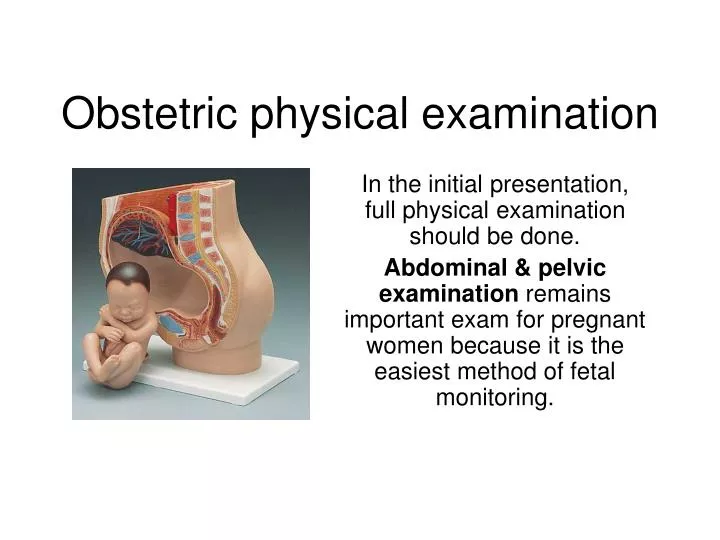
Obstetric physical examination. In the initial presentation, full physical examination should be done. Abdominal & pelvic examination remains important exam for pregnant women because it is the easiest method of fetal monitoring.

E N D
Obstetric physical examination In the initial presentation, full physical examination should be done. Abdominal & pelvic examination remains important exam for pregnant women because it is the easiest method of fetal monitoring.
Essential definitions that you should know to understand the physical examination findings: • The presentation: is the part of the fetus in the lower pole of the uterus overlying the pelvic brim (cephalic, breech)
The lie of the fetus: is the relation of the long axis of the fetus to the uterus (could be longitudinal, oblique or transverse. only longitudinal lie is normal)
The attitude: is the posture of the fetus (flexion, deflexion, extension)
The position: of the baby in relation to the presenting part of the mother’s pelvis. It is expressed according to the denominator which is : • occiput in vertex presentation • sacrum in breech presentation • mentum in face presentation
Station & engagement Station: is the relation of the presenting part to the ischial spine. If the presenting part is at the level of ischial spine, station =0 Engagement: the descent of the biparietal diameter through pelvic brim. If the head is at the level of ischial spine the head must be engaged.
Method of abdominal exam • Inspection: • Size of the uterus: assess • If the length & breadth are both increased multiple pregnancies, polyhydramnios • If the length is increased only large baby • Shape of the uterus: length should be larger than broad this indicates longitudinal lie. But if the uterus is low and broad indicates transverse fetus lie. • Fetal movement • Contour of the abdomen: full bladder may be visible in late pregnancy. Umbilicus may become everted • Skin changes: look for stretch marks, linea nigra, scars that indicates previous surgeries
Method of abdominal exam • Palpation: by Leopold maneuver-4 maneuvers • Palpate the fundus (to determine if it contains breech, head) • By gentle pressure: • if soft consistency/ indefinite outline breech • If hard, smooth, well defined head • Move your fingertips over the fetal mass to determine mobility and sixe • If can’t move independent from the body breech • If moves freely between fingertips head
Lateral palpation: (determine the position of the fetal back and small parts) Hands are placed on each side of the umbilicus. The fetal spine will palpate as firm, flat and linear. The fetal extremities are palpable by their varying contour and movements. The purpose of this maneuver is to determine whether the fetal back is left or right. Method of abdominal exam
Pelvic palpation: 2 maneuvers Grasp the lower poles of the uterus between fingers and thumbs and comment of the size, flexion and mobility of the head. To determine the position of the vertex presentation: try to palpate the prominences (occiput @ the same side of the back & sincipital @ the opposite side of the back) If the sinciput is higher the occiput well flexed If both prominances are at the same level deflexed If can’t palpate the prominances, and the bulk of the head is felt at the same side of the back extended Method of abdominal exam
occiput siniciput)
Method of abdominal exam • Auscultation: help assess fetal well being Auscult the whole abdomen trying to locate the point of maximum intensity • Don’t forget to perform a pelvic exam (details of pelvic exam will be discussed in gynecological exam)but important landmarks to notice during pelvic exam are • Pubis symphasis • Ischial spine
After you examine a pregnant women you should answer the following questions 1. What is the fundal height? It is estimated by centimeters from upper border of the fundus to the pubis symphasis by taping measure. The height of the fundus correlates well with the gestational age especially during the weeks of pregnancy.
After you examine a pregnant women you should answer the following questions 2. lie of the fetus: only longitudinal lie is normal 3. Attitude: normally it is full flexion and every fetal joint is flexed. 4. presentation: normally cephalic 5. position: according to the dominator 6. Is the vertex engaged?
Examination during labor • Palpate uterine contractions • Assessment of the cervix dilatation • 1 finger 1-2 cm dilated • 2 fingers 3-4 cms dilated • 3 fingers 5-6 cms dilated • 4 fingers 7-10 cms dilates
3. Effacement of the cervix: thinning of the cervix (%) or length (cm). The cervix is normally 3-5 cms. If cervix is about 2 cm from external to internal os 50% effaced 50% effaced 100% effaced
4. Consistency of the cervix: soft vs. hard. During labor the cervix becomes soft. 5. Position of the cervix: posterior vs. anterior. During labor the cervix changes from posterior to anterior. 6. Membrane is intact or ruptured: assessed by fluid collection in the vagina