Download
1 / 37
390 likes | 783 Views
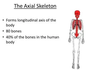
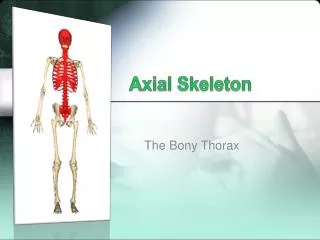
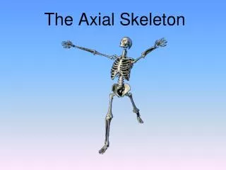
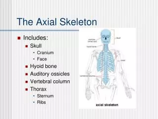
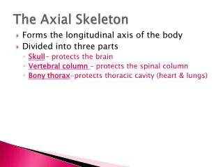
The Axial Skeleton. Forms the longitudinal axis of the body Divided into three parts Skull - protects the brain Vertebral column – protects the spinal column Bony thorax -protects thoracic cavity (heart & lungs). The Axial Skeleton. Figure 5.6a. The Axial Skeleton. Figure 5.6b.

E N D
The Axial Skeleton • Forms the longitudinal axis of the body • Divided into three parts • Skull- protects the brain • Vertebral column – protects the spinal column • Bony thorax-protects thoracic cavity (heart & lungs)
The Axial Skeleton Figure 5.6a
The Axial Skeleton Figure 5.6b
The Skull • Two sets of bones • Cranium • Facial bones • Bones are joined by sutures- interlocking joints; immovable joints that connec bones of skull • Only the mandible is attached by a freely movable joint
Bones of the Cranium • Frontal • Sphenoid • Ethmoid • Right Parietal • Left Parietal • Right Temporal • Left Temporal • Occipital
Facial Bones • Maxillae • Palantine • Zygomatic • Lacrimal • Nasal • Vomer • Inferior Nasal Conchae • Mandible
Human Skull, Lateral View Bone forming anterior cranium Bone pair united by sagittal suture Has greater and lesser wings Site of external auditory meatus Superior and inferior nasal conchae are part of this bone Its “holey plate allows olfactory fibers to pass Allows tear ducts to pass Boney skeleton of the nose Cheek bone Forms most of hard palate Upper jaw Figure 5.7
Human Skull, Superior View • Has greater and lesser wings • Contains a “saddle” that houses the pituitary gland **forms a plateau across the width of the skull Figure 5.8
Human Skull, Inferior View Forms most of hard palate Posterior roof of mouth Inferior part of nasal septum Site of jugular foramen and carotid canal • Its oval-shaped protrusions articulate with the atlas • Spinal cord passes through opening Figure 5.9
Human Skull, Anterior View Sagittal suture Contains a paranasal sinus Contains a paranasal sinus Squamoussutrue (Greater wing) Contains a paranasal sinus • Contain alveoli bearing teeth • Facial bone that contains a sinus Inferior part of nasal septum • Forms the chin • Contain alveoli bearing teeth Figure 5.11
Paranasal Sinuses • Hollow portions of bones surrounding the nasal cavity • Functions of paranasal sinuses • Lighten the skull • Give resonance and amplification to voice
Paranasal Sinuses Figure 5.10a
Paranasal Sinuses Figure 5.10b
The Hyoid Bone *not really a skull bone • The only bone that does not articulate with another bone • Serves as a moveable base for the tongue • Aids in swallowing and speech
The Hyoid Bone Figure 5.12
The Fetal Skull aka BIG HEAD • The fetal skull is large compared to the infant’s total body length • Fetal skull is 1/4th total body length • Adult skull is only 1/8th total body length • Fontanels—fibrous membranes connecting the cranial bones • Allows skull to be compressed during birth and allows for brain growth during late fetal life • Convert to bone within 24 months after birth
The Fetal Skull Figure 5.13a
The Fetal Skull Face is smaller in proportion to cranium Figure 5.13b
The Vertebral Column • Each vertebrae is given a name according to its location • There are 24 single vertebral bones separated by intervertebraldiscs - made up of fibrocartilage • Seven cervical vertebrae are in the neck • Twelvethoracic vertebrae are in the chest region • Fivelumbar vertebrae are associated with the lower back • Herniated disc= a slipped disc; protruding cartilage from vertebra. Causes pain and numbness
The Vertebral Column • Nine vertebrae fuse to form two composite bones • Sacrum- five components; fused • Coccyx- tail bone
The Vertebral Column Figure 5.14
The Vertebral Column • The spine has a normal curvature • Primary curvatures are the spinal curvatures of the thoracic and sacral regions • Present from birth • Secondary curvatures are the spinal curvatures of the cervical and lumbar regions • Develop after birth
The Vertebral Column C shaped spine Figure 5.15
The Vertebral Column Figure 5.16
A Typical Vertebrae, Superior View Figure 5.17
Regional Characteristics of Vertebrae Atlas lacks a body Pivots with C2 Axis articulates with the occipital condyles Figure 5.18a
Regional Characteristics of Vertebrae Forked spinous process Figure 5.18b
Regional Characteristics of Vertebrae Bear facets for articulation with ribs; form part of the bony thoracic cage Figure 5.18c
Regional Characteristics of Vertebrae Vertebrae with blocklike body and short stout spinous process Figure 5.18d
Sacrum and Coccyx • Sacrum • Formed by the fusion of five vertebrae • Forms a joint with the hip bone • Coccyx • Formed from the fusion of three to five vertebrae • “Tailbone,” or remnant of a tail that other vertebrates have
Sacrum and Coccyx Figure 5.19
The Bony Thorax • Forms a cage to protect major organs-cone shaped • Consists of three parts • Sternum • Ribs • True ribs (pairs 1–7) • False ribs (pairs 8–12) • Floating ribs (pairs 11–12) • Thoracic vertebrae
The Bony Thorax Figure 5.20a
Lordosis • Lordosisis a condition that causes the spine to curve towards the body at an exaggerated rate. This curvature makes the individual appear to have a swayback. • Signs of lordosis include a prominent protrusion of the buttocks. An inflexible spine in the affected area signals a severe case of lordosis. Individuals with lordosis and a flexible spine may require no treatment beyond physical therapy. Treatment for lordosis with an inflexible spine includes using a brace and possible surgery.