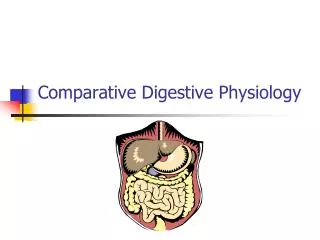
Digestive System: Overview
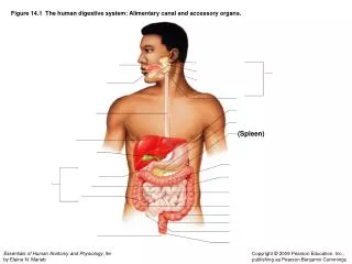
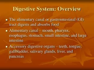
Digestive System: Overview. The alimentary canal or gastrointestinal (GI) tract digests and absorbs food Alimentary canal – mouth, pharynx, esophagus, stomach, small intestine, and large intestine Accessory digestive organs – teeth, tongue, gallbladder, salivary glands, liver, and pancreas.

Digestive System: Overview
E N D
Presentation Transcript
Digestive System: Overview • The alimentary canal or gastrointestinal (GI) tract digests and absorbs food • Alimentary canal – mouth, pharynx, esophagus, stomach, small intestine, and large intestine • Accessory digestive organs – teeth, tongue, gallbladder, salivary glands, liver, and pancreas
Digestive Process • The GI tract is a “disassembly” line • Nutrients become more available to the body in each step • There are six essential activities: • Ingestion, propulsion, and mechanical digestion • Chemical digestion, absorption, and defecation
Gastrointestinal Tract Activities • Ingestion – taking food into the digestive tract • Propulsion – swallowing and peristalsis • Peristalsis – waves of contraction and relaxation of muscles in the organ walls • Mechanical digestion – chewing, mixing, and churning food
Peristalsis and Segmentation Figure 23.3
Gastrointestinal Tract Activities • Chemical digestion – catabolic breakdown of food • Absorption – movement of nutrients from the GI tract to the blood or lymph • Defecation – elimination of indigestible solid wastes
GI Tract • External environment for the digestive process • Regulation of digestion involves: • Mechanical and chemical stimuli – stretch receptors, osmolarity, and presence of substrate in the lumen • Extrinsic control by CNS centers • Intrinsic control by local centers
Receptors of the GI Tract • Mechano- and chemoreceptors respond to: • Stretch, osmolarity, and pH • Presence of substrate, and end products of digestion • They initiate reflexes that: • Activate or inhibit digestive glands • Mix lumen contents and move them along
Nervous Control of the GI Tract • Intrinsic controls • Nerve plexuses near the GI tract initiate short reflexes • Short reflexes are mediated by local enteric plexuses (gut brain) • Extrinsic controls • Long reflexes arising within or outside the GI tract • CNS centers and extrinsic autonomic nerves
Peritoneum and Peritoneal Cavity • Peritoneum – serous membrane of the abdominal cavity • Visceral – covers external surface of most digestive organs • Parietal – lines the body wall • Peritoneal cavity • Lubricates digestive organs • Allows them to slide across one another
Peritoneum and Peritoneal Cavity • Mesentery – double layer of peritoneum that provides: • Vascular and nerve supplies to the viscera • Hold digestive organs in place and store fat • Retroperitoneal organs – organs outside the peritoneum • Peritoneal organs (intraperitoneal) – organs surrounded by peritoneum
Blood Supply: Splanchnic Circulation • Splanchnic- pertaining to the digestive viscera • Arteries and the organs they serve include • The hepatic, splenic, and left gastric: spleen, liver, and stomach • Inferior and superior mesenteric: small and large intestines
Blood Supply: Splanchnic Circulation • Hepatic portal circulation: • Collects nutrient-rich venous blood from the digestive viscera • Delivers this blood to the liver for metabolic processing and storage
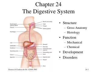
Histology of the Alimentary Canal • From esophagus to the anal canal the walls of the GI tract have the same four tunics • From the lumen outward they are the mucosa, submucosa, muscularis externa, and serosa • Each tunic has a predominant tissue type and a specific digestive function
Mucosa • Moist epithelial layer that lines the lumen of the alimentary canal • Three major functions: • Secretion of mucus • Absorption of end products of digestion • Protection against infectious disease • Consists of three layers: a lining epithelium, lamina propria, and muscularis mucosae
Mucosa: Epithelial Lining • Simple columnar epithelium and mucus-secreting goblet cells • Mucus secretions: • Protect digestive organs from digesting themselves • Ease food along the tract • Stomach and small intestine mucosa contain: • Enzyme-secreting cells • Hormone-secreting cells (making them endocrine and digestive organs)
Mucosa: Lamina Propria and Muscularis Mucosae • Lamina Propria • Loose areolar and reticular connective tissue • Nourishes the epithelium and absorbs nutrients • Contains lymph nodes (part of MALT) important in defense against bacteria • Muscularis mucosae – smooth muscle cells that produce local movements of mucosa
Mucosa: Other Sublayers • Submucosa – dense connective tissue containing elastic fibers, blood and lymphatic vessels, lymph nodes, and nerves • Muscularis externa – responsible for segmentation and peristalsis • Serosa – the protective visceral peritoneum • Replaced by the fibrous adventitia in the esophagus • Retroperitoneal organs have both an adventitia and serosa
Enteric Nervous System • Enteric- pertaining to the intestines • Composed of two major intrinsic nerve plexuses: • Submucosal nerve plexus – regulates glands and smooth muscle in the mucosa • Myenteric nerve plexus – Major nerve supply that controls GI tract mobility • Segmentation and peristalsis are largely automatic involving local reflex arcs • Linked to the CNS via long autonomic reflex arc
Mouth • Oral or buccal cavity: • Is bounded by lips, cheeks, palate, and tongue • Has the oral orifice as its anterior opening • Is continuous with the oropharynx posteriorly
Mouth • To withstand abrasions: • The mouth is lined with stratified squamous epithelium • The gums, hard palate, and dorsum of the tongue are slightly keratinized
Lips and Cheeks • Have a core of skeletal muscles • Lips: orbicularis oris • Cheeks: buccinators • Vestibule – bounded by the lips and cheeks externally, and teeth and gums internally • Oral cavity proper – area that lies within the teeth and gums • Labial frenulum – median fold that joins the internal aspect of each lip to the gum
Palate • Hard palate – underlain by palatine bones and palatine processes of the maxillae • Assists the tongue in chewing • Slightly corrugated on either side of the raphe (midline ridge)
Palate • Soft palate – mobile fold formed mostly of skeletal muscle • Closes off the nasopharynx during swallowing • Uvula projects downward from its free edge • Palatoglossal and palatopharyngeal arches form the borders
Tongue • Occupies the floor of the mouth and fills the oral cavity when mouth is closed • Functions include: • Gripping and repositioning food during chewing • Mixing food with saliva and forming the bolus • Initiation of swallowing, and speech
Tongue • Intrinsic muscles change the shape of the tongue • Extrinsic muscles alter the tongue’s position • Lingual frenulum secures the tongue to the floor of the mouth
Tongue • Superior surface bears three types of papillae • Filiform – give the tongue roughness and provide friction • Fungiform – scattered widely over the tongue and give it a reddish hue • Circumvallate – V-shaped row in back of tongue
Tongue • Sulcus terminalis – groove that separates the tongue into two areas: • Anterior 2/3 residing in the oral cavity • Posterior third residing in the oropharynx
Tongue Figure 23.8
Salivary Glands • Produce and secrete saliva that: • Cleanses the mouth • Moistens and dissolves food chemicals • Aids in bolus formation • Contains enzymes that break down starch
Salivary Glands • Three pairs of extrinsic glands – parotid, submandibular, and sublingual • Intrinsic salivary glands (buccal glands) – scattered throughout the oral mucosa
Salivary Glands • Parotid – lies anterior to the ear between the masseter muscle and skin • Parotid duct opens into the vestibule next to second upper molar • Submandibular – lies along the medial aspect of the mandibular body • Its ducts open at the base of the lingual frenulum • Sublingual – lies anterior to the submandibular gland under the tongue • It opens via 10-12 ducts into the floor of the mouth
Salivary Glands Figure 23.9a
Saliva: Source and Composition • Secreted from serous and mucous cells of salivary glands • 97-99.5% water, hypo-osmotic, slightly acidic solution containing • Electrolytes – Na+, K+, Cl–, PO42–, HCO3– • Digestive enzyme – salivary amylase • Proteins – mucin, lysozyme, defensins, and IgA • Metabolic wastes – urea and uric acid
Control of Salivation • Intrinsic glands keep the mouth moist • Extrinsic salivary glands secrete serous, enzyme-rich saliva in response to: • Ingested food which stimulates chemoreceptors and pressoreceptors • The thought of food • Strong sympathetic stimulation inhibits salivation and results in dry mouth
Teeth • Primary and permanent dentitions have formed by age 21 • Primary – 20 deciduous teeth that erupt at intervals between 6 and 24 months • Permanent – enlarge and develop causing the root of deciduous teeth to be resorbed and fall out between the ages of 6 and 12 years • All but the third molars have erupted by the end of adolescence • Usually 32 permanent teeth
Deciduous Teeth Figure 23.10.1
Permanent Teeth Figure 23.10.2
Classification of Teeth • Teeth are classified according to their shape and function • Incisors – chisel-shaped teeth for cutting or nipping • Canines – fanglike teeth that tear or pierce • Premolars (bicuspids) and molars – have broad crowns with rounded tips; best suited for grinding or crushing • During chewing, upper and lower molars lock together generating crushing force
Dental Formula: Permanent Teeth • A shorthand way of indicating the number and relative position of teeth • Written as ratio of upper to lower teeth for the mouth • Primary: 2I (incisors), 1C (canine), 2M (molars) • Permanent: 2I, 1C, 2PM (premolars), 3M
Tooth Structure • Two main regions – crown and the root • Crown – exposed part of the tooth above the gingiva • Enamel – acellular, brittle material composed of calcium salts and hydroxyapatite crystals; the hardest substance in the body • Encapsules the crown of the tooth • Root – portion of the tooth embedded in the jawbone
Tooth Structure • Neck – constriction where the crown and root come together • Cementum – calcified connective tissue • Covers the root • Attaches it to the periodontal ligament
Tooth Structure • Periodontal ligament • Anchors the tooth in the alveolus of the jaw • Forms the fibrous joint called a gomaphosis • Gingival sulcus – depression where the gingiva borders the tooth
Tooth Structure • Dentin – bonelike material deep to the enamel cap that forms the bulk of the tooth • Pulp cavity – cavity surrounded by dentin that contains pulp • Pulp – connective tissue, blood vessels, and nerves
Tooth Structure • Root canal – portion of the pulp cavity that extends into the root • Apical foramen – proximal opening to the root canal • Odontoblasts – secrete and maintain dentin throughout life
Tooth and Gum Disease • Dental caries – gradual demineralization of enamel and dentin by bacterial action • Dental plaque, a film of sugar, bacteria, and mouth debris, adheres to teeth • Acid produced by the bacteria in the plaque dissolves calcium salts • Without these salts, organic matter is digested by proteolytic enzymes • Daily flossing and brushing help prevent caries by removing forming plaque
Tooth and Gum Disease: Periodontitis • Gingivitis – as plaque accumulates, it calcifies and forms calculus, or tartar • Accumulation of calculus: • Disrupts the seal between the gingivae and the teeth • Puts the gums at risk for infection • Periodontitis – serious gum disease resulting from an immune response • Immune system attacks intruders as well as body tissues, carving pockets around the teeth and dissolving bone