Download

1 / 32
420 likes | 1.42k Views
Chapter 8 Special Senses. Special senses Smell Taste Sight Hearing Equilibrium. The Senses. General senses of touch Temperature Pressure Pain. The Eye and Vision. 70 % of sensory receptors are in eyes Each eye has over a million nerve fibers Protection for the eye - bony orbit

E N D
Special senses Smell Taste Sight Hearing Equilibrium The Senses General senses of touch • Temperature • Pressure • Pain
The Eye and Vision • 70 % of sensory receptors are in eyes • Each eye has over a million nerve fibers • Protection for the eye - bony orbit - surrounding fat
Accessory Structures of the Eye • Eyelids • Eyelashes • Meibomian glands • modified sebaceous glands • oily secretion to lubricate • Ciliary glands Modified sweat glands between eyelashes • Conjunctiva - Membrane lining eyelids; connects to eye surface; secretes mucus to lubricate
Accessory Structures of the Eye Lacrimal apparatus • Lacrimal gland – produces lacrimal fluid • Lacrimal canals – drains lacrimal fluid from eyes • Lacrimal sac – provides passage of lacrimal fluid towards nasal cavity • Nasolacrimal duct – empties lacrimal fluid into the nasal cavity
Function of the Lacrimal Apparatus • Properties of lacrimal fluid - Dilute salt solution (tears) - Contains antibodies and lysozymes • Protects, moistens, & lubricates the eye • Empties into the nasal cavity
Extrinsic Eye Muscles • Muscles attach to the outer surface of the eye • Produce eye movements Figure 8.2
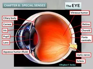
Structure of the Eye The wall is composed of three tunics • Fibrous tunic – outside layer • Choroid – middle layer • Sensory tunic –inside layer Figure 8.3a
The Fibrous Tunic • Sclera • White connective tissue layer • Seen anteriorly as the “white of the eye” • Cornea • Transparent, central anterior portion • Allows for light to pass through • Repairs itself easily • The only human tissue that can be transplanted without fear of rejection
Choroid Layer • Blood-rich nutritive tunic • Pigment prevents light from scattering • Modified interiorly into two structures • Cilliary body – smooth muscle • Iris • Pigmented layer that gives eye color • Pupil – rounded opening in the iris
Sensory Tunic (Retina) • Contains receptor cells (photoreceptors) • Rods • Cones • Signals pass from photoreceptors via a two-neuron chain • Bipolar neurons • Ganglion cells • Signals leave the retina toward the brain through the optic nerve
Neurons of the Retina Figure 8.4
Neurons of the Retina and Vision Rods • Most are found towards the edges of the retina • Allow dim light vision and peripheral vision • Perception is all in gray tones Cones • Allow for detailed color vision • Densest in the center of the retina • Fovea centralis – area of the retina with only cones Optic disk (Blind spot)- No photoreceptor cells
Cone Sensitivity • There are three types of cones • Different cones are sensitive to different wavelengths • Color blindness is the result of lack of one cone type Figure 8.6
Lens • Biconvex crystal-like structure • Held in place by a suspensory ligament attached to the ciliary body
Internal Eye Chamber Fluids Aqueous humor - Watery fluid in chamber between lens & cornea • Similar to blood plasma • Helps maintain intraocular pressure • Provides nutrients for the lens and cornea • Reabsorbed into blood by the canal of Schlemm Vitreous humor - Gel-like substance behind lens • Keeps the eye from collapsing • Lasts a lifetime and is not replaced
Lens Accommodation • Light must be focused to a point on the retina for optimal vision • eye is set for distance vision (over 20 ft away) • lens must change shape to focus for closer objects
Images Formed on the Retina Figure 8.10
Visual Pathway • Photoreceptors of retina • Optic nerve • Optic nerve crosses at the optic chiasma • Optic tracts • Thalamus (axons form optic radiation) • Visula cortex of the occipital lobe
Eye Reflexes • Internal muscles controlled by autonomic nervous system - Bright light causes pupils to constrict (radial and ciliary muscles) • Viewing close objects causes accommodation • External muscles control eye movement to follow objects • Viewing close objects causes convergence (eyes moving medially)
lens which has become opaque or clouded Cataracts Causes? Diabetes, old age, pollution?
visual defect in which the eyes are misaligned and point in different directions misalignment of the eyes STRABISMUS
disease of the eye in which damage occurs to the optic nerve, typically as a result of an elevated pressure within the eye. . Damage to the optic nerve causes progressive loss in peripheral vision and can eventually lead to blindness. GLAUCOMA
wedge-shaped fibrovascular growth of conjunctiva that extends onto the cornea benign lesions that can be found on either side of the cornea. PTERYGIUM
irregularity in the shape of the cornea or the lens. Instead of being shaped round, the cornea is shaped oval, causing a blurred image at all distances. Patients may notice blurred or ghost images close up or far away. present in various degrees Astigmatism
(Farsightedness) -unable to see near objects without extreme focusing. images are formed behind the retina eye too short, or the refractive powers of cornea & lens are too weak Hyperopia
(Nearsightedness)-Distant objects are unclear in cases of myopia. condition of the eye in which images are formed in front of the retina the eye is relatively too long or refractive powers of the cornea & lens are too strong. Myopia
Corneal degenerative disorder Cornea becomes progressively thin and steep The front of the eye bulges. Keratoconus
“Pink eye” Infection of conjuctiva Caused by bacteria or virus Highly contagious Conjunctivitis
Complimentary Colors Stare at the flag for 30 seconds. Then look at a white surface. What happens & why?